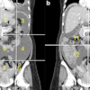
| FIGURE 1.3.3 Side branch IPMN. Coronal reformatted CT image (A) shows cystic dilatation of a single side branch in the pancreatic head (arrow). On subsequent ERCP (B), contrast opacifies the same dilated side branch. Coronal T2-weighted SSFSE MR image (C) from a second patient shows a cluster of cystic lesions in the pancreatic head, which on MRCP (D) appears as a branching multicystic structure that communicates with, but does not directly involve, the main pancreatic duct. Contrast-enhanced CT (E) and intraoperative US (F) images from a third patient show a more nonspecific cystic lesion involving the pancreatic tail. Contrast-enhanced CT (G), T2-weighted MR (H), and MRCP (I) images from a final patient show a multicystic-appearing side branch IPMN involving the uncinate process. |
Atlas of Gastrointestinal Imaging Figure 1.3.3 Side branch IPMN
Latest in Home


Sponsored
Turning Innovation into Everyday Efficiency
April 22, 2026
Lu-177 PSMA-SPECT/CT predicts survival in mCRPC
April 29, 2026


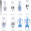
![A 53-year-old patient (patient number four) with a recurrent pituitary adenoma with extension of a cystic component of disease to the medial temporal lobe apparent on MRI (contoured in blue), and extension of disease to the left sphenoid bone and orbital apex apparent on [68Ga]Ga-DOTA-TATE (contoured in yellow).](https://img.auntminnie.com/mindful/smg/workspaces/default/uploads/2026/04/pituitary-tumor.QGsEnyB4bU.jpg?auto=format%2Ccompress&dpr=2&fit=crop&h=167&q=70&w=250)


