Minnies finalists, page 3
Scientific Paper of the Year
The concept of gaslighting in the radiology workplace: Concepts and implications. Ballane B et al, American Roentgen Ray Society annual meeting, 2021. Learn more about this study.

The first study up for the Scientific Paper of the Year shined a light on the concept of gaslighting in radiology -- that is, manipulating co-workers or patients into doubting their experiences or shutting down their knowledge or expertise.
In a presentation at the virtual American Roentgen Ray Society meeting in April, a team led by Dr. Brandon Ballane of New York Medical College in Valhalla, NY, discussed how a workplace's atmosphere can dramatically influence the effectiveness of its staff and why abuse such as gaslighting can be so damaging.
The team described how to recognize gaslighters: They tend to manipulate colleagues to gain power in the relationship, often via guilt, and promote a false narrative about colleagues to other members of the workplace.
What's the best way to counter gaslighting? The team suggested addressing medical staff's concerns in a calm, conversational manner and empowering members of the radiology staff to make collaborative diagnostic decisions can help prevent gaslighting.
Ultimately, when gaslighting behavior is identified, the "victim" should speak to the person directly, as well as to fellow staff members to determine whether the behavior is isolated or widespread, according to Ballane and colleagues.
COVID-19 vaccination-associated axillary adenopathy: Imaging findings and follow-up recommendations in 23 women. Mortazavi S, American Journal of Roentgenology, February 24, 2021. Learn more about this study.
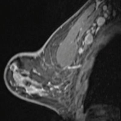 Breast MRI after first COVID-19 vaccination dose. Image courtesy of the American Roentgen Ray Society.
Radiology continues to play a pivotal role in responding to the COVID-19 crisis, as the second finalist for Scientific Paper of the Year showed by warning that vaccines can manifest on imaging in ways that mimic disease.
Dr. Shabnam Mortazavi of the University of California, Los Angeles called for practice recommendations to prevent excessive follow-up imaging and potential biopsy of COVID-19 vaccination-associated axillary adenopathy.
In the study of 23 women who underwent breast imaging after being vaccinated, researchers found that most of the adenopathy on breast imaging occurred with the Pfizer vaccine (52%), among asymptomatic women presenting for either screening or diagnostic purposes (86%), and on ultrasound (52%).
Incorporating a patient's COVID-19 vaccination history, including vaccination date and laterality, is critical to optimize assessment and management of imaging-detected axillary adenopathy in women with otherwise normal breast imaging, Mortazavi stated.
Warnings aside, this report and others with similar findings concluded with messages encouraging patients to get vaccinations.
Best New Radiology Device
Two of radiology's leading modalities -- CT and MRI -- and two of its largest vendors -- Siemens Healthineers and Philips Healthcare -- face off in the final round of this year's Minnies competition in the Best New Radiology Device category.
Magnetom Free.Max MRI scanner, Siemens Healthineers
First introduced in November 2020, Magnetom Free.Max is the product of a Siemens effort to develop an MRI scanner that's based on superconducting technology but that has many of the benefits of a permanent magnet system, such as easier siting and operating costs.
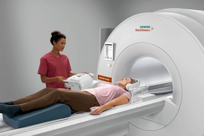 Siemens' Magnetom Free.Max 0.55-tesla MRI scanner. Image courtesy of Siemens Healthineers.
First off, the 0.55-tesla scanner needs just 1 L of helium to keep its magnet cold, and the system doesn't require a quench pipe. And the scanner's small size makes it easy to install in a wider variety of locations -- many of which can be closer to patients than the radiology department.
Siemens is also touting the 80-cm bore of the scanner, which makes the system well-suited to scanning claustrophobic or obese patients. Magnetom Free.Max uses the same interface as other scanners in the company's Magnetom product line, and it's also available with Siemens' myExam Companion technology, which helps radiologic technologists set up and perform exams with standardized protocols.
Free.Max also supports Deep Resolve, a set of algorithms that perform targeted denoising and utilize deep learning to provide sharper, higher-resolution images. Siemens announced U.S. Food and Drug Administration (FDA) clearance for Free.Max in July 2021.
Spectral CT 7500 spectral CT scanner, Philips Healthcare
Spectral CT (also known as dual-energy CT) is based on a simple principle: Cancers and healthy tissue respond differently to x-ray beams sent at different energy levels. Up until now, however, actually performing spectral CT on a routine clinical basis has not been so simple. The technology can require a different workflow compared with conventional CT, and it tends to impart a slightly higher radiation dose.
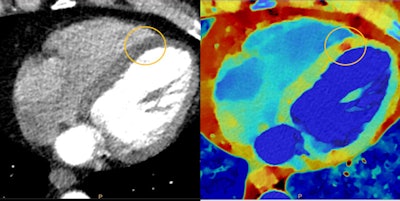 Myocardial perfusion defects are difficult to visualize on conventional CT (left) due to beam-hardening artifacts, but they are visible on spectral CT (right). Image courtesy of Philips Healthcare.
Philips tackled these issues head-on with Spectral CT 7500. Launched in May 2021, the scanner is designed to make spectral CT easy enough to perform for routine daily use. The system acquires 8 cm of data per gantry rotation, which eliminates the need for image stitching. Philips also reduced the number of mouse clicks required to generate spectral images.
Radiation dose was addressed with an enhanced detector and imaging chain that allows single-pass image acquisition at 100 kVp, compared with 120 kVp for the previous generation of Philips spectral CT scanners.
Spectral CT 7500 can operate in spectral mode for a variety of clinical applications, including cardiology, oncology, neurology, and trauma. The company believes that spectral CT can save healthcare costs by eliminating the need to perform follow-up studies after an inconclusive exam.
The scanner has both 510(k) clearance in the U.S. and the CE Mark in Europe.
Best New Radiology Software
AI computer-assisted triage software for incidental detection of pulmonary embolism on CT, Aidoc
Computer-aided triage has emerged over the last few years as perhaps the most popular artificial intelligence (AI) application in radiology, so it's not surprising that Aidoc's AI software for incidental detection of pulmonary embolism (PE) on CT exams has been voted in as a finalist for Best New Radiology Software.
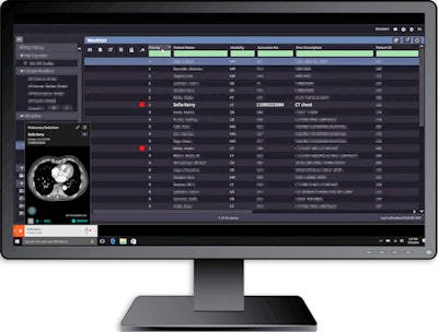 AI computer-assisted triage software for incidental detection of pulmonary embolism on CT. Image courtesy of Aidoc.
In contrast to Aidoc's algorithm for detecting PE on CT pulmonary angiography exams -- the 2019 Minnie winner for Best New Radiology Software -- this computer-aided triage algorithm flags and communicates incidentally detected findings of PE on nondedicated exams. It's available for use with CT scanners from Philips Healthcare and Siemens Healthineers.
The algorithm received FDA clearance in October 2020.
Genius AI software for breast tomosynthesis, Hologic
Our other finalist for Best New Radiology Software is designed to highlight suspicious areas of interest on digital breast tomosynthesis exams to aid radiologists in interpretation. Integrated into Hologic's Dimensions platform, Genius AI also provides important workflow benefits, according to the vendor.
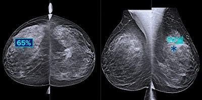 Hologic's Genius AI software highlights suspicious areas of interest on breast tomosynthesis exams. Image courtesy of Hologic.
Running on the acquisition workstation of the mammography system without the need for a server, the deep-learning software delivers key metrics to radiologists to help them categorize and prioritize cases by complexity and expected reading time, according to the vendor. Hologic said studies have shown that Genius AI is more accurate than its previous-generation computer-aided detection software.
Hologic garnered FDA clearance for Genius AI in late 2020.
Best New Radiology Vendor
Prescient Imaging
A developer of portable PET scanning technology, Prescient Imaging made headlines earlier this year with news that its BBX-PET portable PET scanner had secured FDA 510(k) clearance.
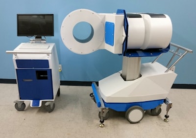 Prescient Imaging's BBX-PET mobile PET scanner with workstation. Image courtesy of Prescient Imaging.
Over seven years in the making, BBX-PET is designed to be wheeled to the point of care and can provide PET imaging of the brain, breast, and extremities, according to the vendor. BBX-PET is suitable for use by hospitals and imaging centers, as well as neurology, geriatric, psychiatry, and radiation oncology centers, according to co-founder and CEO Farhad Daghighian, PhD.
Applications include research, diagnosis, therapeutic planning, and therapeutic outcome assessment. Over $8 million was invested by Daghighian and private investors in developing BBX-PET, the company said.
Prescient is also working on several other PET systems including VH-PET -- a PET scanner that's designed to be added to existing CT or MRI scanners and can be used in vertical or horizontal mode. In addition, Biopsy PET Dx is a PET scanner targeted at imaging of small tissue samples, while P-Arm aims to support interventional molecular imaging applications.
Rad AI
Founded by radiologists, Rad AI is focused on utilizing artificial intelligence technology to streamline workflow and deliver significant daily time savings for radiologists.
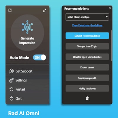 Rad AI's Omni software for automated radiology report impressions. Image courtesy of Rad AI.
The firm's initial offering -- Omni -- automatically generates the radiology report's impression section in a manner that's customized to the preferred language of each radiologist. The radiologist then reviews and signs off on the report. The result is an overall increase in productivity of up to 25% -- or over 60 minutes -- each day, according to Rad AI.
Rad AI has also developed Continuity, an application that's designed to automatically close the loop on significant incidental findings, according to the company.
The company was officially launched in late 2019 with help from a $4 million seed round led by Google's artificial intelligence-focused venture fund Gradient Ventures.
Best Educational Mobile App
Radiology Assistant 2.0, BestApps (iOS)
A perennial contender for Best Educational Mobile App, Radiology Assistant 2.0 has been named a finalist for the fifth time. It also previously won the award in 2018, and its predecessor, Radiology Assistant, was a finalist in 2013.
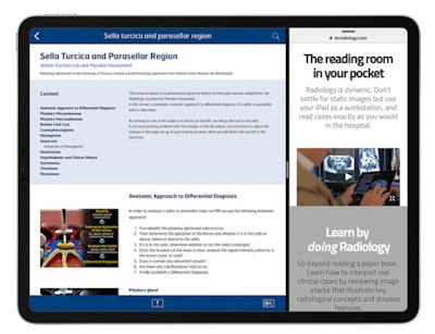 Radiology Assistant 2.0. Image courtesy of Dr. Wouter Veldhuis, PhD.
Developed by Dutch radiologists Dr. Wouter Veldhuis, PhD, of the University Medical Center Utrecht and Dr. Robin Smithuis of Rijnland Hospital Leiderdorp, Radiology Assistant 2.0 features peer-reviewed articles written by expert radiologists. Smithuis is the founder of the Radiology Assistant educational website and nonprofit organization.
Many new articles have been added recently to Radiology Assistant 2.0, both to help radiologists read COVID-19 scans, as well as to learn about important topics such as uterine anomalies, lung fibrosis, gallstone disease, ankle fractures, Crohn's disease, and breast ultrasound, according to Veldhuis.
The app is free to download, but it requires a subscription to access article categories. It also offers in-app purchases, supports full-text searching in all articles, and allows users to view high-resolution versions of images in full-screen mode without leaving their place in the article.
RadDiscord, a server on Discord, Dr. Grace Zhu (iOS, Android)
Launched in October 2020 as a server on the Discord platform to provide a community for radiology residents to collectively study for the radiology board exam, RadDiscord aims to facilitate real-time interactions for its members from around the world.
The server -- #RadDiscord -- features board review lectures, Anki flashcards, study voice channels, and text channels with case-of-the-day and subject content, according to RadDiscord.
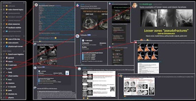 RadDiscord educational content created by radiology residents. Image courtesy of Dr. Grace Zhu.
In addition to its goal of providing equal access to radiology knowledge to all practicing radiologists and trainees, RadDiscord also aims to continuously improve how radiologists are educated. RadDiscord was created by Dr. Grace Zhu, a former radiology resident and future faculty member at the University of Utah.
Best Radiology Image
The two finalists in the Best Radiology Image category pit a PET scan of a patient who showed abnormal uptake after getting a COVID-19 vaccine against a series of images demonstrating the power of AI-based image reconstruction for MRI. The finalists were selected through voting on AuntMinnie.com's Facebook page.
The image below is from a study published May 20 in the American Journal of Roentgenology and is one of a number of case reports and studies published this year that show how vaccination for COVID-19 can cause pathology that appears to look like cancer.
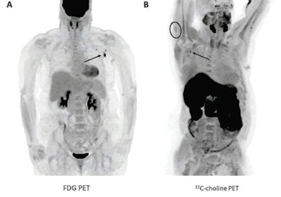 PET imaging showed abnormal radiotracer uptake in the lymph nodes of cancer patients after they received COVID-19 vaccinations. (A) A 57-year-old woman with right upper arm melanoma who received the first dose of the COVID-19 vaccine (Pfizer-BioNTech) in the left deltoid 15 days prior to FDG PET/CT. FDG uptake is observed within left axillary lymph nodes (arrow, SUVmax = 9.3). (B) A 62-year-old man with metastatic prostate carcinoma who received the second dose of COVID-19 vaccine (Pfizer-BioNTech) in the right deltoid seven days prior to C-11 choline PET/CT. Image courtesy of the American Journal of Roentgenology.
The second finalist in the Best Radiology Image category comes from a presentation at the European Congress of Radiology in March 2021 by Dr. Suzie Bash, a neuroradiologist at image services provider RadNet. The image series demonstrates how AI can be used to speed up MRI data reconstruction times while maintaining image quality.
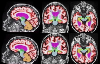 AI-based image reconstruction enables significantly faster brain MRI scan times while maintaining quantification accuracy and enhancing image quality. Representative 3D T1-weighted multiplanar images with volumetric segmentation on a 3-tesla scanner. (Left to right): Sagittal, coronal, and axial T1-weighted images with standard protocols (scan time, 5 minutes and 44 seconds) on the top row and deep learning-enhanced accelerated imaging (scan time, 2 minutes and 18 seconds) on bottom row. Images courtesy of Dr. Suzie Bash.
Previous page | 1 | 2 | 3


