Dear Advanced Visualization Insider,
We have a lot to learn about disease processes in the lung, but a team led by University of Iowa researchers believes it has developed a method that may improve our understanding.
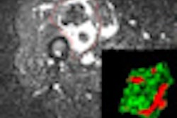
Employing microCT imaging and 3D image reconstruction tools, the group has produced 3D renderings of the pulmonary acinus in mice. With the ability to image the actual structure of the most peripheral respiratory units of the lung, the researchers hope to gain new insight into the lung's response to inhaled pollutants, for example, or to model new delivery systems for inhaled drugs.
Our coverage of the research is the subject of this newsletter's Insider Exclusive. You can access the article before our other members by clicking here.
In other news in the Advanced Visualization Digital Community, find out how vascular neurologists at the Mayo Clinic in Arizona are using the iPhone for emergency stroke evaluations.
In addition, a Japanese team recently determined that the combination of an advanced iterative reconstruction technique with 3D processing might offer considerable value in both reduced- and low-dose CT scans for patients with pulmonary diseases. Both image quality and interreader agreement improved after the method was applied. For the details, click here.
Computer-aided detection (CAD) software can be a valuable tool for radiologists, but it's still important that readers not focus their attention primarily on areas of images with CAD marks, a group from Brigham and Women's Hospital and Harvard Medical School concluded. Click here to find out why.
We're also featuring a profile of the 3D lab at Massachusetts General Hospital. Read about the secrets to its success.
Do you have any interesting images or clips that might be suitable for our AV Gallery? You are welcome to submit them here.


