Dear Advanced Visualization Insider,
It can be challenging to diagnose fibrosing interstitial lung diseases, even for expert multidisciplinary discussion teams. But quantitative analysis of texture patterns on high-resolution CT exams can yield better survival predictions in these patients, according to this edition's Insider Exclusive.
Researchers from the University of Pennsylvania found that a texture-based image classification model substantially outperformed expert radiologist diagnostic labels of usual interstitial pneumonia versus nonusual interstitial pneumonia for predicting patient survival.
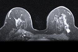
Radiomics can also improve the accuracy of breast MRI in differentiating malignant from benign axillary lymph nodes in women with breast cancer, according to a new study. Researchers from the University of Cincinnati Medical Center found that textural image analysis enables radiologists to obtain quantitative information that may not be visible to the human eye, which could help clinicians develop more detailed axillary management plans for patients.
Another recent study reported that two radiomics features on low-dose CT exams in lung cancer screening can be used to identify early-stage lung cancer patients who may be at higher risk for poor survival outcomes, potentially enabling earlier interventions.
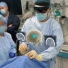
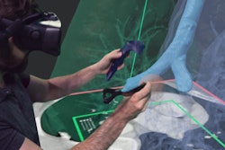
The future is now for 3D imaging technologies such as cinematic rendering, 3D printing, simulation training, virtual reality, and augmented reality, according to Dr. Ramin Javan of George Washington University Hospital. Speaking of augmented reality, a group from the Cleveland Clinic recently described the technology's potential for improving the speed and safety of liver tumor ablation procedures.
Can 3D printing help improve the performance of ultrasound elastography? Researchers from Germany found that a 3D-printed device with built-in sensors could produce more accurate shear-wave elastography measurements for breast imaging and other use cases.
A team from the University of Wisconsin has concluded that cardiac 4D flow MRI can quantify blood flow in the ascending aorta and main pulmonary artery during strenuous exercise, offering potential as a tool for evaluating right-sided heart dysfunction. Also, a 3D software algorithm can identify small changes in knee joints over time on MRI exams. And measurements of muscle mass and density automatically extracted from chest CT exams can be predictive of all-cause mortality over a six-year period in older men.
Do you have an idea for a story you'd like to see covered in the Advanced Visualization Community? Please feel free to drop me a line.


