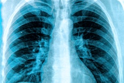
The use of artificial intelligence (AI) software in daily clinical practice for detecting lung lesions on chest x-rays has been an overall positive experience, according to researchers from a large hospital in South Korea.
A team led by Dr. Hyun Joo Shin, PhD, of Yongin Severance Hospital in Yongin, surveyed radiologists and clinicians at their hospital two years after implementing commercially available software (Insight CXR, Lunit) as an aid in detecting lung lesions on chest x-rays. Respondents said the tool was most useful in the emergency room, and pneumothorax was considered the most valuable finding, according to the findings.
"It is notable that many doctors now routinely refer to AI in their everyday workflow as this gives us a glimpse of what full adaptation of AI can mean for radiology in the future," the group wrote in an article published March 2 in PLOS One.
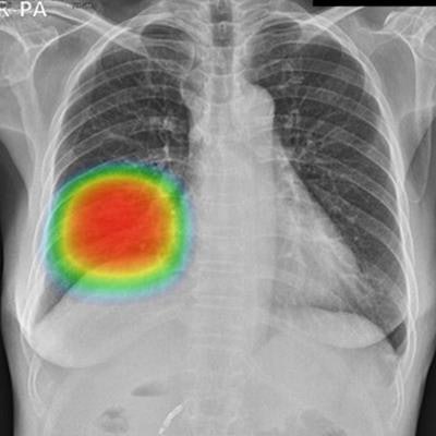
Lunit's Insight CXR was approved in Korea in 2018. The software indicates the location of lesions suspicious for major lung abnormalities, such as nodules, consolidation, and pneumothorax.
The software has been available for use at Yongin Severance Hospital since March 2020 as an AI-based aid for interpreting chest images. In this study, the researchers aimed to find out what doctors think about the actual integration of the software in their daily practice.
The group sent a hospital-wide online survey in 2021 to all clinicians and radiologists (194 total) and received 123 responses, with 74% completing all questions. Questions were designed to gather information on basic demographics, experience with AI, actual individual use, and preferences and attitudes toward AI-based software.
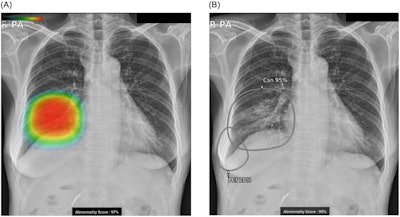 Chest radiographs of a 57-year-old female with pneumonia and right pleural effusion. Images are results analyzed with (A) version 2 and (B) version 3 of the AI-based lesion detection software. (A) Version 2 can detect and display three types of lesions (consolidation, nodule, and pneumothorax) with a color heatmap and total abnormality score. (B) Version 3 can detect and display nine types of lesions (six additional types of the lesion in addition to the three lesions detected in version 2) with a grayscale heatmap and abnormality score for each lesion. Note the right pleural effusion that was additionally detected and displayed with version 3 of the software. Image courtesy of PLOS One through CC BY 4.0.
Analysis of the results showed that the proportion of individuals who used the AI program was higher among radiologists than clinicians (83% vs. 46%, p = 0.008), with AI perceived as being the most useful in the emergency room, and pneumothorax considered the most valuable finding.
About 21% of clinicians and 16% of radiologists changed their own reading results after referring to the AI program, the group reported. The most common reason for using the software was to reduce missed diagnoses, and the second reason was that the software made it easy to utilize the AI results on PACS, they wrote.
In addition, trust levels for AI were 64.9% among clinicians and 66.5% among radiologists. The participants reported that AI helped reduce reading times and reading requests, the researchers wrote.
"Interestingly, 28% of participants answered that referring to the AI results had become routine during their readings of chest radiographs," the group noted.
Since the survey was sent in 2021, it is possible that the perceptions of doctors about AI have since changed and further continuous studies are warranted as experience accumulates, the researchers wrote.
"We are in the process of demonstrating the effect of AI on diagnostic accuracy or reading time to justify these survey results in an objective way and hope to confirm our findings with quantitative results in the next step of our research," the group concluded.

