A cross-sectional brain MRI study comparing white matter hemodynamics suggests that the hormone oestrogen may play a neuroprotective role in the aging process, according to one in a series of Young Investigator Award presentations Monday at the International Society for Magnetic Resonance in Medicine (ISMRM) meeting.
The retrospective study based at the Athinoula A. Martinos Center for Biomedical Imaging in Boston is part of the Human Connectome Project, a worldwide effort to explore how age, sex, disease, and other factors can affect the ever-changing connections in the human brain. For this white-matter investigation, the researchers measured cerebral blood flow (CBF), arterial transit time (ATT), and fractional anisotropy (FA) in over 600 healthy participants between 35 and 90 years of age (evenly split between males and females) across four sites in the U.S.
Data processing was extensive, said presenter Nikou Louise Damestani, PhD, formerly of Harvard Medical School, Massachusetts General Hospital, and Kings College London. Damestani said the study used two pipelines, one for diffusion imaging, the other for perfusion imaging. Tract-based spatial statistics were helpful in this early validation of white matter hemodynamics as a potential biomarker preceding structural decline and cognitive change in the brain.
"We don't know very much about what happens to white matter hemodynamics during the aging process, largely because the studies that have been done so far have lacked spatial specificity, and the studies that have been done, that have tried to probe this, have technical limitations and require advanced processing techniques that can be difficult to implement for white matter," Damestani explained.
While much is known about the brain's gray matter and white matter structure, there have been inconsistent findings as to what happens to white matter hemodynamics during the aging process. It is also unknown how hemodynamics correlates with structural decline or if it could be a useful biomarker for characterizing the aging process.
For potential validation, Damestani and colleagues have taken the first step, measuring white matter hemodynamics with spatial specificity in a large, healthy adult cohort. To that end, they produced an FA skeleton, CBF skeleton, and ATT skeleton for every individual in the study.
The researchers used two high-resolution approaches. One is a technique called multidelay pseudocontinuous arterial spin labeling (md-pCASL), or labeling blood at the level of the neck and allowing the blood to perfuse to different regions of the brain, allowing different post-labeling delay times, with a series of times allowed for the perfusion to take place. When they subtracted the labeled and unlabeled images and applied a quantification process, they were able to extract the CBF measure. Multidelay pCASL also quantified the ATT or the time taken for blood to reach the brain tissue from the labeling plane.
The other technique using 3-tesla diffusion imaging produced 1.5 millimeter (mm) isotropic resolution.
Damestani and colleagues developed their quantification approach for CBF and ATT in-house and then projected the corresponding maps onto the white matter skeletons.
The framework for this study complemented gray matter studies by identifying the relationship between white matter hemodynamics and age and sex. Damestani said the team mapped CBF and ATT on average across the entire cohort within white matter, with the spatial specificity they were looking for.
"Interestingly, we found substantial overlap for lower average CBF and higher average ATT in the posterior circulation. This could be linked to the known biomechanical changes that take place in the posterior cerebral arteries during the aging process and ultimately indicates that CBF and ATT are complementary hemodynamic measures. They're not measuring the exact same thing. We're seeing the perfusion rates once the blood reaches the tissue and the time it takes for the blood to reach that region."
Another finding confirmed that lower FA is associated with older age and longer ATT across the vast majority of white matter. There were regions where lower CBF was associated with older age, but in other regions, this didn't exist, Damestani continued.
"When we probed this further, we found that this could be because ATT has a strong linear relationship whereas CBF has a more nonlinear relationship with age," she said.
Finally, with regard to sex, lower ATT was observed globally for females compared to males. Higher CBF in females showed more localized findings in which the differences were predominantly in the posterior regions. For FA, the researchers found it was lower in females in the corticospinal tract, and this has been found in previous studies. CBF and ATT had much bigger differences than FA at a tract level.
"The differences in physiology could be linked to hormonal differences that are inherent to men and women as they age," Damestani said. "For example there are some studies suggesting that oestrogen has a neuroprotective role and so we really recommend considering this in your aging research."
The team already has preliminary findings looking at the menopause transition as well, which were not shared during the presentation.
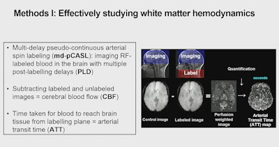 Young Investigator Award finalist Nikou Louise Damestani, PhD, explained a study of white matter hemodynamics. The work raises questions about white matter hemodynamics as a potential biomarker preceding structural decline and cognitive change in the brain.
Overall, toward correlating structural integrity and hemodynamics, Damestani said ATT (rather than CBF) may be a more sensitive measure in structural decline because ATT can capture the delay that takes place due to breakdowns seen with aging, for example as a result of arterial stiffening. Longitudinal studies will help to validate the team's findings.
"There's one big missing link for aging research, which is a biomarker that precedes structural decline and cognitive change," Damestani said. "We also need a biomarker that can predict structural decline more effectively than some of the gray matter measures.
"In theory, white matter hemodynamics could be the target," Damestani said. "What we need to know next is at what point do white matter changes start occurring in the physiology that could be linked to structural decline that we know takes place."
Other ISMRM 2024 Young Investigator Award finalists:
Retta El Sayed, PhD, from the Georgia Institute of Technology in Atlanta, for "Assessment of Complex Flow Patterns in Patients with Carotid Webs, Patients with Carotid Atherosclerosis and Healthy Subjects Using 4D Flow MRI." The team quantified parameters related to vascular dysfunction, including wall shear stress (WSS) and oscillatory shear index (OSI). The results showed that subjects with carotid webs have larger regions of complex blood flow represented by low WSS and high OSI. The study improves understanding of disturbed hemodynamics caused by carotid webs in comparison to atherosclerosis, which may explain the mechanism of thrombus formation and lay the groundwork for stroke risk assessment in patients with carotid webs, according to Sayed and colleagues.
Shohei Fujita, MD, PhD, from Juntendo University in Tokyo, Japan, for "Cross-Vendor Multiparametric Mapping of the Human Brain Using 3D-QALAS: A Multicenter and Multivendor Study." This study addressed unmet need for a cross-vendor, multiparametric technique to facilitate data pooling across sites. Intrascanner repeatability and intervendor reproducibility were evaluated in vivo on five different 3-tesla (3T) systems from four vendors (GE HealthCare, Philips, Siemens Healthineers, and Canon Medical Systems). Fujita and colleagues evaluated a vendor-standardized multiparametric mapping scheme based on 3D-QALAS for whole-brain T1, T2, and proton density (PD) mapping. 3D-QALAS provided T1, T2, and PD with coefficient of variations < 4% using 3T scanners from different manufacturers.
Victor Han, PhD, from the University of California at Berkeley, for "Any-nucleus Distributed Active Programmable Transmit (ADAPT) Coil." The ADAPT Coil appears to enable arbitrary nucleus excitation in high-field MRI and may reduce the cost and technological barriers of clinical translation of X-nuclei research, according to Han and colleagues.
Jessie Mosso, PhD, from CIBM Center for Biomedical Imaging in Lausanne, Switzerland, for "Diffusion-Weighted SPECIAL Improves the Detection of J-Coupled Metabolites at Ultrahigh Magnetic Field." Mosso and team proposed their new sequence for single-voxel diffusion-weighted hydrogen 1 (1H) MR spectroscopy, called DW-SPECIAL, as an alternative to STE-LASER when strongly J-coupled metabolites like glutamine are investigated, thereby extending the range of accessible metabolites in the context of diffusion-weighted MRS acquisitions at high field.
Aviad Rabinowich, MD, from Tel Aviv University in Israel, for "Fetal MRI-Based Body and Adioposity Quantification for Small for Gestational Age Perinatal Risk Stratification." Rabinowich and colleagues aimed to stratify perinatal risk using MRI-based body composition metrics. Quantifying fetal body composition through MRI can offer additional insights into the severity of small for gestational age complicated pregnancies, according to the researchers.
Felicia Seemann, PhD, from the National Institutes of Health, for "Dynamic Lung Water Magnetic Resonance Imaging During Exercise Stress." Quantification of lung water during exercise is of interest for early diagnosis of heart failure. Current clinical tests to assess lung water include qualitative chest x-rays, semiquantitative lung ultrasound, and invasive right heart catheterizations, none of which provide 3D resolution. However, MRI is promising for quantifying lung water at rest and shortly after exercise, Seemann said. Seemann and team developed a method that uses continuous 10-minute 3D lung MRI acquisition with a sliding window and motion-corrected image reconstruction to quantify transient lung water dynamics between rest and exercise stress. The code is available open source at github.com/NHLBI-MR/dynamic_lung_water. The next step in this research is to image patients before and after diuretics and eventually over time with other pharmacological interventions.
Rui Tian from Max Planck Institute for Biological Cybernetics in Germany, for "Accelerated 2D Cartesian MRI with an 8-channel local B0 Coil Array Combined with Parallel Imaging." This team investigated accelerated MRI with flexible modulations of nonlinear B0 fields using a custom-built local B0 array. The experiment was conducted on a Siemens 9.4-tesla human MRI scanner. The B0 coil array allows for independent control over each local coil elements a variety of nonlinear views can be synthesized to allow flexible, spatially varying nonlinear phased modulation on an object, according to Tian. Among other findings, the MRI sampling theory is extended to rigorously compare distinct nonlinear field encoding schemes in a quantitative k-space. The proposed field calibration technique substantially enhances reconstruction, given flexible modulations of local B0 coils, Tian said.

