CAPE TOWN - MR and MR spectroscopy (MRS) imaging reveal how exercising and better nutrition can improve metabolic measures in obese patients, according to a talk delivered May 12 at the International Society of Magnetic Resonance in Medicine (ISMRM) meeting.
The improved measures include liver fat, visceral adiposity, intramuscular lipid accumulation, and skeletal muscle mitochondrial function, said Martin Krššák, PhD, of the Medical University of Vienna, to session attendees.
"This study demonstrates the power of combined multi-organ MRI/MRS to noninvasively monitor metabolic adaptations to lifestyle intervention in obesity, providing novel biomarkers of improved lipid metabolism and mitochondrial function that may guide personalized obesity management and metabolic health strategies," he said.
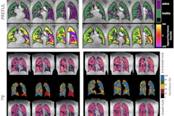
Obesity changes energy metabolism across a range of organs, Krššák noted, but data regarding the effects of lifestyle interventions are limited. To address this knowledge gap, he and colleagues conducted a study that included 51 middle-aged adults with obesity (mean body mass index [BMI], 34 kg/m). Of these, 36 participants made up an intervention group and completed three supervised aerobic and strength-training sessions weekly alongside nutritional counseling and psychological support over 12 weeks. Fifteen individuals served as controls.
All study participants underwent 3T and 7T MRI/MRS before and after the intervention, and the team tracked liver and adipose fat composition as well as the patients' muscles strength. The intervention program consisted of three sessions of supervised aerobic and strength training, weekly nutritional counseling, and monthly psychological support.
Using both 3T and 7T MR and MR spectroscopy imaging on the same day, Krššák and colleagues noted the metabolics of three types of tissue: liver, adipose, and the gastrocnemius muscle. This approach allowed them to simultaneously assess lipid content and fatty acid composition in the liver and fat depots while also tracking muscle performance during exercise.
 Martin Krššák, PhD
The investigators found that after three months, the intervention group showed significant reductions in waist circumference (p = 0.02), hip circumference (p = 0.01), and BMI (p = 0.01). There were no significant changes in the control group.
They also reported the following:
- Liver fat content declined significantly (p = 0.01), as did intramyocellular fat deposits in the gastrocnemius (p = 0.001).
- Visceral fat volume decreased (p = 0.04).
- Fatty acid composition of visceral adipose tissue shifted toward greater unsaturation (p = 0.004) -- a change associated with improved metabolic flexibility.
Skeletal muscle mitochondrial capacity showed meaningful gains, indicating enhanced aerobic energy production following exercise training, Krššák reported.
Results of a three-month intervention on obese patients | ||
Measure | Before intervention | After intervention |
| Waist circumference (cm) | 106.8 | 98.2 |
| Hip circumference (cm) | 120.7 | 110.8 |
| BMI (kg/m2) | 35 | 32.2 |
| Percent fat | 43.1 | 38.7 |
| Percent muscle | 25.9 | 28 |
The findings underscore the potential of advanced MR techniques to offer sensitive, noninvasive biomarkers for tracking metabolic responses to obesity treatment, Krššák said. Because the assessment is noninvasive and covers multiple organ systems in a single session, the approach could guide individualized treatment decisions, helping to identify which patients show early hepatic or mitochondrial improvement and which may need a further-modified program.
"As 7T systems become more accessible, this integrated imaging approach could translate into a practical tool for metabolic phenotyping in clinical obesity programs," Krššák concluded.
Check out AuntMinnie’s ShowCast for full coverage of ISMRM 2026.