Dear Cardiac Imaging Insider,
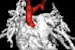
Early reports on the use of 320-detector-row CT for cardiac imaging are promising. First, the ability to scan the whole heart in less than a beat may permit reduced contrast dose and more uniform opacification of the coronary arteries. The radiation dose is dropping, too. Last month, researchers from Brigham and Women's Hospital in Boston found that prospectively gated 320-detector-row CT angiography studies could be acquired with radiation doses of less than 5 mSv.
Today we bring you a report from Nevada Imaging Center in Las Vegas, which has been scanning heart patients with 320-detector-row CT for about six months. Doctors there are producing high-quality, low-dose cardiac studies even in patients with fast heartbeats and profound arrhythmias (though not necessarily both at the same time).
In this era of tightly controlled healthcare costs, they believe cardiac CT is a promising solution to the need for faster, cheaper workup of patients with suspected heart disease. Get the rest of the story in this issue's Insider Exclusive, brought to you before it's posted for other AuntMinnie.com members.
Dual-source CT has also jumped on the low-dose bandwagon. A new study from Switzerland reports extremely low doses in step-and-shoot mode. Of course, if you want a side of functional assessment with that cardiac CT special, you'll still need retrospective electrocardiogram gating, ramping up radiation to approximately 10 mSv in most scanners using tube current modulation.
Studies continue to show reduced costs with CT as well. A new analytic model lends further support to the notion that cardiac CT may be cheaper than the standard of care.
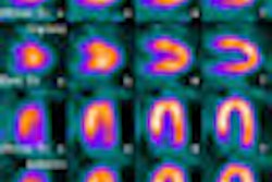
Meanwhile, nuclear medicine proves its enduring relevance with a new study from Durham, NC. Duke researchers found an association between impaired myocardial perfusion and sudden cardiac death, and SPECT myocardial perfusion imaging also lent incremental prognostic information to clinical and other measures of cardiac function, the group reported.
At the American Society of Nuclear Cardiology (ASNC) meeting earlier this month, a multinational team reported that images from a new ultrafast cardiac gamma camera were comparable to those of standard dual-detector cameras, potentially reducing radiation dose to the patient. Staff writer Wayne Forrest reports on the promising new technology here.
Also at the ASNC meeting, a proof-of-concept study showed how FDG-PET/CT might have a role in detecting and evaluating coronary plaque.
In ultrasound, real-time 3D transesophageal echocardiography was used to guide the treatment of patients with heart conditions such as atrial septal defect and congenital heart disease.
Be sure to scroll through the stories below for more news in cardiac imaging, or visit your Cardiac Imaging Digital Community.


