Dear Orthopedic Insider,
MRI is supplanting other modalities -- particularly plain radiography -- for a variety of musculoskeletal imaging applications. But orthopedic imaging specialists may encounter challenges when shifting from x-ray to MRI due to differences in the way that musculoskeletal anatomy appears on each modality.
The concordance -- or lack of same -- between x-ray and MRI in imaging patients with hip dysplasia was the subject of research presented at the International Society for Magnetic Resonance in Medicine (ISMRM) meeting in Seattle. Staff writer Shalmali Pal was on hand to report on the presentation, which is the subject of this edition's Insider Exclusive.
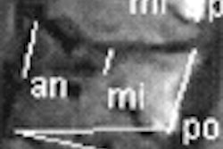
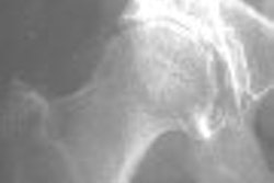
In the study, the researchers compared MRI and radiography measurements of key elements of hip anatomy in patients with developmental dysplasia of the hip (DDH). They found that MRI and x-ray measurements agreed in only one of four categories used to measure hip dysplasia. Find out the implications of their research for patient management by clicking here.
In another article we're featuring this week, we profile a pair of studies that examined whether cholesterol levels and antioxidants can have a meaningful impact on bone health, as measured on bone mineral density (BMD) exams. Learn about that study by clicking here.
Meanwhile, researchers at the Hospital for Special Surgery in New York City are using ultrasound to guide injections of cortisone for treating De Quervain's tendonitis, a painful repetitive strain injury, while Spanish researchers have found that BMD decreases during the first two years of androgen deprivation therapy (ADT) in men with prostate cancer.
Get these stories and more by visiting the Orthopedic Imaging Digital Community, at orthopedic.auntminnie.com, and if you have any ideas or suggestions for stories, please feel free to e-mail me at [email protected].