Endobronchial Carcinoid
The patient shown below was a 27 year old male with complaints of recurrent pulmonary infection in the right middle lobe.
(Click the small images to view the larger radiographs)
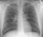
The PA chest radiograph revealed obscuration of the right heart border suggesting
infiltrate:
The lateral view, however, was more suspicious for right middle lobe atelectasis.
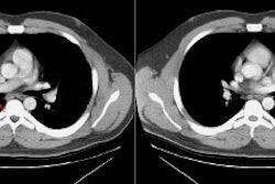
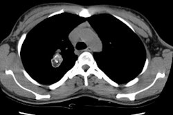
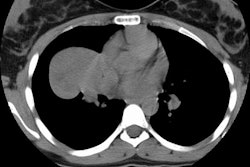
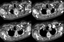
A CT scan was performed due to the chronic nature of the patients complaints. The CT
scan demonstrated a filling defect within the right middle lobe bronchus (red arrows) with
post-obstructive atelectasis and probably some mucous plugging within the distal bronchi.
Unfortunately, submucosal extension of the tumor to the interlobar bronchus was found
histologically, and the patient was treated with a bilobectomy.
Soft tissue windows are also provided:
