Sclerosing Hemangioma:
View cases of sclerosing hemangioma
Clinical:
Sclerosing hemangioma is a rare benign tumor of the lung [2]. The tumor is felt to be primarily a proliferation of epithelial cells, probably type II pneumocytes, although the exact histiogenesis is uncertain [1]. About 80% of patients with the lesion are female [1]. Although considered a benign lesion, it can manifest rarely as multiple tumors, as a tumor nodule surrounded by multiple daughter nodules in the same lobe, or with hilar lymph node metastases (these findings are generally associated with large lesions) [2].
X-ray:
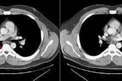
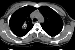
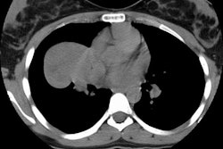
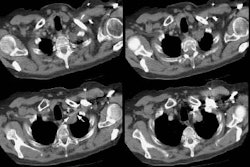
On CXR, the lesion appears as a well-defined rounded or oval mass [2]. On CT the lesion appears as a well circumscribed, homogeneous, enhancing soft tissue mass [2]. The lesion may contain areas of decreased attenuation consistent with necrosis. Calcification can be seen in about one-third of cases [1,2]. The lesion is frequently juxta-pleural and can be located within the fissures.
REFERENCES:
(1) J Comput Assist Tomogr 1994; Im JG, et al. Sclerosing hemangiomas of the lung and interlobar fissures: CT findings. 18: 34-38
(2) AJR 2006; Chung MJ, et al. Pulmonary sclerosing hemangioma presenting as a solitary pulmonary nodule: dynamic CT findings and histopathologic comparisons. 187: 430-437


