AJR Am J Roentgenol 1995 Jan;164(1):57-61. Bronchogenic carcinoma in HIV-positive patients: findings on chest radiographs and CT scans.
Fishman JE, Schwartz DS, Sais GJ, Flores MR, Sridhar KS
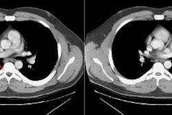
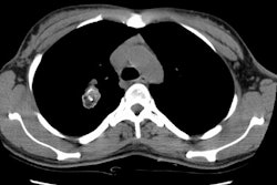
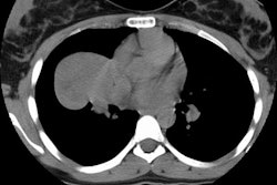
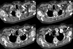
OBJECTIVE. The radiographic manifestations of bronchogenic carcinoma in HIV-positive individuals may resemble or accompany changes of inflammatory disease. To provide information that is useful in the differential diagnosis, we studied the findings on plain radiographs and chest CT scans in 30 HIV-positive patients with proven bronchogenic carcinoma and correlated the radiographic features with the presence or absence of thoracic opportunistic infection. SUBJECTS AND METHODS. Thirty HIV-positive individuals had bronchogenic carcinoma diagnosed at our institution between 1986 and 1993. Fourteen (47%) of the 30 had AIDS at the time of cancer diagnosis. All but one of the patients were men, and the median age at diagnosis was 48 years (range, 32-66 years). Most (90%) had a history of smoking. Eighteen (60%) of the 30 had a history of pulmonary tuberculosis, Pneumocystis carinii pneumonia, or both. We retrospectively reviewed all available chest radiographs (n = 27) and chest CT scans (n = 25) for tumor size and location, adenopathy, pleural disease, and pulmonary infiltrates. RESULTS. Eighteen tumors (60%) were peripheral, 11 (37%) were central (hilar or mediastinal), and one manifested as a metastatic pleural mass. Of the peripheral tumors, 17 (94%) were in the upper lobes. All the central tumors showed obstructive consolidation of lung in the distribution of the affected airway. Adenopathy was present in 63% of the patients, and pleural effusions or masses were seen in 33%. A history of tuberculosis or Pneumocystis carinii pneumonia was present in 83% of the patients with peripheral tumors but only 27% of the patients with central lesions (p = .005). Superimposed infiltrates were present in six patients (20%). Three (17%) of 18 peripheral tumors were obscured by or mistaken for inflammatory disease, delaying the diagnosis of cancer. CONCLUSION. Bronchogenic carcinoma usually manifests as a peripheral upper lobe mass in HIV-positive patients with a history of tuberculosis or Pneumocystis carinii pneumonia, whereas central masses are more common in patients without a history of thoracic opportunistic infection. Carcinoma should be suspected in patients with peripheral lesions that persist despite appropriate antibiotic therapy.
PMID: 7998569, MUID: 95091216


