Thoracic carcinoids: radiologic-pathologic correlation.
Rosado de Christenson ML, Abbott GF, Kirejczyk WM, Galvin JR, Travis WD.
Department of Radiologic Pathology, Armed Forces Institute of Pathology,
Washington, DC 20306-6000, USA.
Carcinoids are neuroendocrine neoplasms. Bronchial carcinoids are unusual,
malignant primary neoplasms that characteristically involve the central airways
and typically exhibit well-defined margins and bronchial-related growth.
Bronchial carcinoids include low-grade typical carcinoids and the more
aggressive atypical carcinoids. These tumors usually affect patients in the 3rd
through 7th decades of life who are often symptomatic with cough, hemoptysis, or
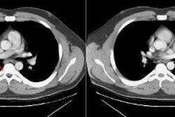
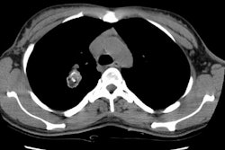
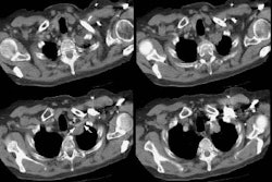
obstructive pneumonia. Bronchial carcinoids radiologically manifest as hilar or
perihilar masses, with or without associated atelectasis, pneumonia,
bronchiectasis, or mucoid impaction. At computed tomography, an anatomic
relationship of these tumors to a bronchus is usually seen, and they may show
contrast material enhancement or calcification. In rare cases, carcinoids occur
in the thymus; when they do, they are aggressive tumors that affect adults who
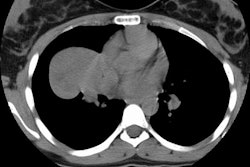
usually present with chest pain, cough, and dyspnea. Thymic carcinoids manifest
radiologically as anterior mediastinal masses and may mimic thymomas. Thoracic
carcinoids are treated by surgical excision. The prognosis for patients with
typical bronchial carcinoids is excellent; patients with atypical bronchial or
thymic carcinoids have a worse prognosis.
Tumor > Malignant > Carcinoid
Radiographics 1999 May-Jun;19(3):707-36
Latest in Tumor
Tumor > Benign > Inflammatory myofibroblastic tumor
October 19, 2020
Tumor > Malignant > Lungcancer > Staging
February 22, 2009
Tumor > Benign > Mesenchymal hamartoma
October 30, 2008
Tumor > Benign > Leiomyoma
October 30, 2008


