Mucoepidermoid Carcinoma:
Clinical:
A rare (0.1-0.2% of all pulmonary malignancies [3]), locally invasive tumor, most commonly located in the lobar or segmental bronchi, and uncommonly in the trachea or main bronchi. The tumor is believed to originate from the minor salivary glands lining the tracheobronchial tree [3]. Up to 25-28% of patients can be asymptomatic [1]. Symptoms include cough, dyspnea, wheezing, hemoptysis, and chest pain. Although the tumor can occur at any age, about half of patients are between the ages of 30-40 years, and between 18-50% occur in patients under the age of 30 [3,4].Histologically the lesion is classified as either low or high grade depending on the amount of cellular atypia, mitotic activity, local invasion, and necrosis [4]. The METC1-MAML2 fusion oncogene is present in 75-100% of mucoepidermoid carcinomas [5]. Low grade tumors tend to be located in the central bronchi and igh grade lesions tend to be located in the peripheral lung [4]. Metastases to local nodes is rare in patients with low grade lesions (0-2%), but can be found in up to 15% of patient with high grade lesions. Low grade lesions have a very good prognosis.
X-ray:
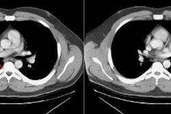
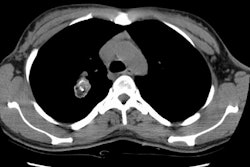
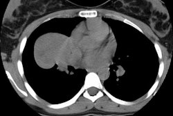
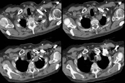
The CXR will usually reveal post-obstructive pneumonia or atelectasis.On CT the mass appears as a smooth surfaced, enhancing, endobronchial mass (usually in a main or lobar bronchus) which conforms to the bronching airway [3,4,5]. A lobular heterogeneous lesion containing multilobular cystic structures filled with low attenuation fluid has also been described [4]. High grade lesions may have a more ragged, invasive appearance with necrosis. Mucous secretion in high grade tumors is decreased due to lack of differentiation and cystic lesions are less common [4]. Punctate calcification can be seen in up to 50% of lesions [1]. FDG uptake is commonly seen with high and intermediate grade tumors [2].
REFERENCES:
(1) Radiology 1999; Kim TS, et al. Mucoepidermoid carcinoma of the
tracheobronchial tree: radiographic and CT findings in 12
patients. 212: 643-648
(2) AJR 2007; Jeong SY, et al. Integrated PET/CT of salivary gland type carcinoma of the lung in 12 patients. 189: 1407-1413
(3) Radiographics 2009; Park CM, et al. Tumors in the
tracheobronchial tree: CT and FDG PET features. 29: 55-71
(4) AJR 2015; Wang YQ, et al. Low-grade and high-grade
mucoepidermoid carcinoma of the lung: findings and clinical
features of 17 cases. 205: 1160-1166
(5) Radiographics 2018; Lichtenberger JP, et al. Primary lung tumors in children: radiologic-pathologic correlation. 38: 2151-2172


