Carcinoid Case 2:
The patient was an active duty male troop who presented with compliants of chronic cough.
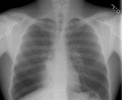
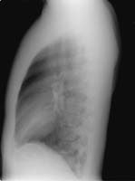
The chest radiograph revealed a right infrahilar density and increased retrocardiac density on the right (yellow arrows). The was hyperlucency laterally within the right lower lobe and the findings were felt to be suspicious for right lower lobe volume loss. The lateral exam is also provided by was unrevealing.
(Click on small images to view larger radiographs)
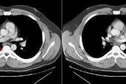
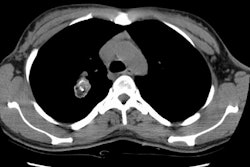
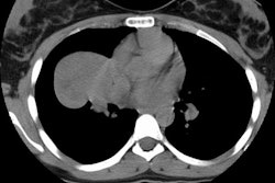
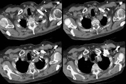
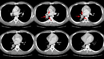
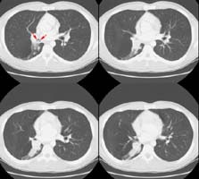
A CT scan of the chest was performed and revealed a heterogeneous soft tissue mass in the right interlobar bronchus with post-obstructive atelectasis.
A photo from the patient's bronchoscopy revealed the intralumenal mass. The mass proved to be a typical carcinoid or type I Kulchitsky cell carcinomas.

