Hemangioma:
Clinical:
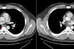
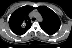
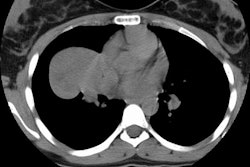
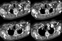
The capillary hemangioma is the most common tracheal tumor in the neonate. The lesion is most commonly found in the subglottic airway and girls are affected twice as often as boys. Patients are usually not symptomatic at birth, but over 90% of cases become symptomatic by age 6 months. The lesion will typically grow for a 12-18 month period, and then slowly involute typically by age 2 to 3 years (however, the involution can be variable and last up to 10 years [2]). Patients typically have intermittent stridor which may be exacerbated by crying, feeding, or exercise. Close interval follow-up or laser ablation can be used to treat these patients. The lesion typically appears as a purple, blue, or pinkish soft-tissue mass arising from the posterior wall of the trachea. The lesion may occasionally be anterior or circumferential. On plain films, a hemagioma appears as a sharply marginated, soft tissue mass within the subglottic trachea. On CT, there is intense enhancement of the lesion with contrast [2].Pulmonary hemangioma are uncommon. The most common presenting symptom is hemoptysis. The lesion appears as a solitary pulmonary nodule generally peripheral, in a subpleural location.
REFERENCES:
(1) J Thorac Imag 1995; 10: 180-198 (p. 184-185)
(2) AJR 2005; Koplewitz BZ, et al. CT of hemangioma of the upper airways in children. 184: 663-670


