Metastatic Breast Cancer
The patient was a 44 year old female who presented with a 10 cm fungating left breast mass.
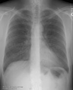
The initial chest radiograph demonstrated multiple small nodular densities in the lungs. The soft tissue defect from the breast carcinoma is best appreciated on the lateral view, but some soft tissue asymmetry is also evident on the PA exam. (Click on the small images to view the larger radiographs)
PA: 
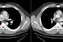
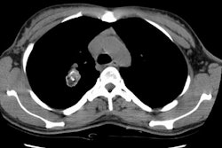
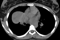
A CT scan of the chest demonstrated the large, cavitary left breast mass, the presence of pathologic adenopathy in the left axilla, and multiple pulmonary metastases.
Soft tissue: 


