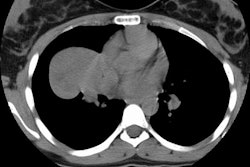
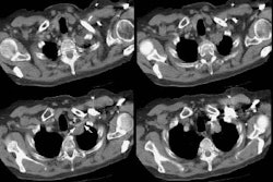
Atypical Carcinoid
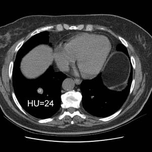
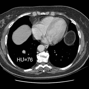
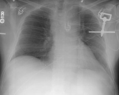
The patient was a middle aged female who had a slowly enlarging mass in the right lower lung. The patient was reluctant to have surgery, but contrast enhanced CT revealed nodule enhancement greater than 20 HU which suggested a malignant lesion and the patient agreed to surgery. The lesion proved to be a carcinoid tumor with atypical features at histologic analysis. Peripheral carcinoids are more frequently atypical as in this case.CXR revealed a right lower lobe non-calcified mass (yellow arrow):
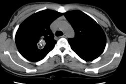
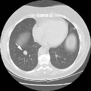
The lesion had well defined margins at CT (white arrow):
The lesion enhanced 52 HU after IV contrast administration which suggested a malignant lesion- it was removed and found to be an atypical carcinoid:
