The usefulness of bone scintigraphy with SPECT images for detection of pulmonary metastases from osteosarcoma.
Pevarski DJ, Drane WE, Scarborough MT
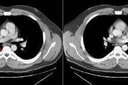
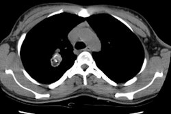
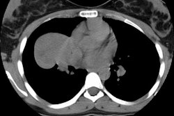
OBJECTIVE: We prospectively compared the ability of two techniques--bone scintigraphy with single-photon emission computed tomography (SPECT) of the chest and CT of the chest--to reveal potential osteosarcoma metastases of the lung. SUBJECTS AND METHODS: Our study included 27 patients with osteosarcoma who prospectively underwent both bone scintigraphy with SPECT of the chest and CT of the chest. The imaging results were compared with outcome or pathologic analysis of any lung lesions found. RESULTS: Eight (30%) of the 27 patients had pulmonary metastases. Four of these eight patients had positive results on both CT studies and bone SPECT studies, with additional lesions detected with bone SPECT in two of these four patients. The other four patients with pulmonary metastases had positive results on CT studies, whereas the results of bone SPECT studies remained negative. The results of bone SPECT studies were negative in the 19 patients without pulmonary metastases. CT, however, showed abnormalities in seven (37%) of the 19 patients, which were eventually attributed to benign conditions. CONCLUSION: Negative results on a bone SPECT study of the chest should not be used to exclude the possibility of lung metastases. However, if the results are positive, a bone SPECT study can be used to confirm abnormalities seen on CT scans and may also reveal subtle lesions missed on CT scans.


