Ann Thorac Surg 1999 Mar;67(3):790-7
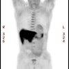
Evaluation of fluorine-18-fluorodeoxyglucose whole body positron emission
tomography imaging in the staging of lung cancer.
Saunders CA, Dussek JE, O'Doherty MJ, Maisey MN.
BACKGROUND: Surgical resection of lung cancer remains the treatment of choice in
appropriately staged disease, but conventional imaging techniques have
limitations. Positron emission tomography (PET) may improve staging accuracy.
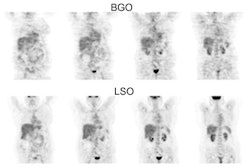
METHODS: We studied whole body and localized thoracic PET in staging lung
cancer. Standardized uptake value was calculated for the primary lesion.
Ninety-seven patients under consideration for surgical resection were included.
PET, computed tomography, and clinical staging were compared to stage at
operation, biopsy, or final outcome. Mean follow up was 17.5 months. RESULTS:
PET detected all primary lung cancers with two false-positive primary sites.
Sensitivity and specificity for N2 and N3 mediastinal disease was 20% and 89.9%
for computed tomography and 70.6% and 97% for PET. PET correctly altered stage
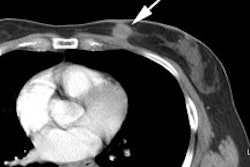
in 26.8%, nodal stage in 13.4%, and detected distant metastases in 16.5%. PET
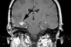
missed 7 of 10 cerebral metastases. PET altered management in 37% of patients.
PET staging (p<0.0001) and standardized uptake value (p<0.001) were the
best predictors of time to death apart from operative staging. CONCLUSIONS: PET
provides significant staging and prognostic information in lung cancer patients
considered operable by standard criteria. Routine use of PET will prevent
unnecessary operation and may be cost effective.
