Developing and testing a new class of “super phantoms” is needed to optimize new medical imaging techniques before they are used in human studies, according to an article published May 24 in Communications Engineering.
An international group from the U.S., the Netherlands, and Germany, said there is a major knowledge gap between testing on current phantoms and testing on highly complex human tissues. Super phantoms may bridge this gap, according to the authors, and they provided illustrated examples, as well as addressed development challenges.
“We call these models ‘super phantoms’ to signify that they occupy a class of physical and biological realism beyond standard imaging phantoms,” noted lead author Srirang Manohar, PhD, of the University of Twente in Enschede, the Netherlands.
Phantoms are test objects used for initial testing and optimization of medical imaging techniques. In the face of rapidly emerging imaging technology, however, standard phantoms have not kept pace, as they rarely capture the complex properties of human tissue, the authors wrote.
In their article, the group introduced the concept of super phantoms, offering the following definition: “A computer model, an inanimate physical object, or an ex vivo organ that replicates the most relevant anatomic and functional imaging biomarkers of the structure of interest under carefully controlled laboratory settings.”
Recently developed examples include a digital four-dimensional (4D) CT breast imaging super phantom. This super phantom includes not only the morphological representation of breast tissues and tumors, but also incorporates the wash-in and wash-out kinetics of iodinated contrast-containing blood in vasculature and simulated tumors, the authors wrote.
Such a super phantom could be used in 4D imaging using dedicated breast CT to explore the perfusion properties of tumors for optimized treatment, the group wrote.
 Examples of test objects that the authors classify as super phantoms. Asterisks indicate those presented in the article, with reference numbers provided in square brackets and the imaging modality for which the super phantom is intended in round brackets. The drawing of the body is inspired by the Vitruvian man created by Leonardo Da Vinci in 1490.Image courtesy of Communications Engineering.
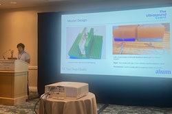
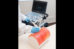
Other examples include a physical photoacoustic-ultrasound imaging super phantom that accurately represents the main tissues found in the female breast, including skin, fat, and fibroglandular tissue, and an ex vivo 4D flow MRI super phantom, which researchers could use to investigate the impact of various surgical procedures on intra-cardiac flow and evaluate the performance of MRI sequences in different pathophysiological settings, the authors wrote.
Concrete steps to integrate these new models in research and development include establishing a super phantom core facility, the group noted. A centralized shared facility would cater to multiple institutions and be equipped with high-quality fabrication technology and dedicated personnel.
“This facility would ideally be located at a university with a strong biomedical engineering program and established collaborations with academic hospitals, to house a range of equipment and supplies,” the group wrote.
Ultimately, methods to develop super phantoms should be akin to those used in achieving human space flight, where a focus on rigorous testing and optimization on earth using specialized life-sized simulators and training modules was key, the group wrote.
“We advocate for a comparable emphasis and commitment to rigorous laboratory testing during the development of advanced medical imaging technologies, prior to human testing,” the authors concluded.
The full article is available here.


