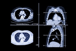
SAN FRANCISCO - Compared with other obstructive pulmonary diseases, the course of emphysema remains a bit hazy. Even among heavy smokers, just 40% will develop severe emphysema, say presenters at the Chest 2000 conference this week. But CT scanning has shed new light on the disease by quantifying its severity and determining which patients would benefit from lung volume reduction surgery (LVRS).
"Not all patients with chronic obstructive pulmonary disease (COPD) or chronic bronchitis have emphysema. Why should clinicians really be concerned about it?" said Dr. Robert Rogers from the University of Pittsburgh. "Where we thought that emphysema was a fairly hopeless disease, I think we now have some new interventions -- LVRS, retinoic acids -- that mean we have to find newer ways to quantify the severity of the disease."
Rogers reviewed the results of two studies he co-authored that used CT to judge the severity of emphysema. The first paper, published in 1999 in the American Journal of Respiratory and Critical Care Medicine, used CT to assess lung surface area in patients with emphysema.
"The question we asked was ‘Can we better interpret CT of the lung and select better candidates for LVRS?’ We wanted to do a quantitative analysis and eliminate subjective visual analysis," he explained.
In this study, non-contrast, 10-mm thick CT slices were obtained with a 9800 HiLight Advantage scanner (GE Medical Systems, Waukesha, WI) from patients with mild and severe emphysema, as well as a control group, one week before surgery.
According to the results, the CT estimates of lung volume showed a higher total lung and airspace volume in patients with severe emphysema. Subjects in this same group had less tissue volume and lower lung weight. With the CT scans, the clinicians were able to track the destruction of the lung surface area by emphysema, as well as provide specific locations where the lung had been destroyed by the disease (AJRCCM, March 1999, Vol.159:3, pp.851-856).
"The exciting thing about these measurements is that it’s going to give us a way to see if we are restoring surface area [through intervention]," Rogers said.
Rogers added that the group chose to use conventional CT rather than high-resolution CT in order to obtain the maximum amount of information about lung volume.
"The problem with high-resolution CT is that you are looking at 1-mm depth and you are ignoring all the space in between. We think that is a loss of data. In a 10-mm slice, we are doing the entire volume of the lung, whereas high resolution ignores about 85% of lung volume," he said.
In a second study scheduled to appear in the November issue of Chest, Rogers and his colleagues used CT to prospectively evaluate candidates for LVRS. The comparison of baseline CT and post-operative scans showed that total lung volume was reduced from 6666.74 ml/g to 5778.83 ml/g in treated patients.
Future research will focus on examining the lungs’ inner and outer cores with CT to see if clinicians can improve on their ability to predict the success of LVRS, Rogers said.
"The strengths of our method are that we are able to quantify the entire volume of the lung, and it will be easily reproduced in a radiology department in the future," he said.
CT will play a role in several ongoing studies that will assess new emphysema-fighting pharmaceuticals, according to presenter Dr. Frank Sciurba from the University of Pittsburgh. In a trial funded by the National Institutes of Health called the Feasibility of Retinoid Therapy in Emphysema (FORTE), CT scans are providing investigators with changes in lung density and texture after treatment with retinoic acid.
By Shalmali PalAuntMinnie.com staff writer
October 26, 2000
Let AuntMinnie.com know what you think about this story.
Copyright © 2000 AuntMinnie.com


