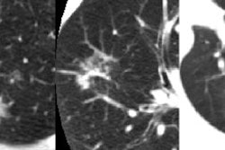
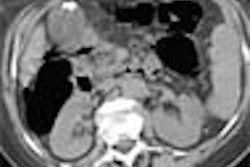
Indirect CT venography (CTV) and duplex venous sonography turn in similar performance for evaluating acute deep vein thrombosis (DVT) in ICU patients with suspected pulmonary embolism, according to an article in the August American Journal of Roentgenology.
"CT pulmonary angiography with CTV offers a single first-line diagnostic test in the ICU for evaluation of suspected DVT that offers high accuracy in diagnosing pulmonary embolism and results comparable to those of lower extremity venous sonography," wrote Dr. Matthew Taffoni and colleagues from the Medical University of South Carolina (MUSC) in Charleston, SC. "Therefore, good-quality CTV obviates sonography. In equivocal cases or with poor venous opacification, confirmation with sonography is still recommended."
To prospectively compare the two approaches, the MUSC research team studied all patients undergoing CT pulmonary angiography in the evaluation of acute pulmonary embolism between June 2002 and April 2003 (AJR, August 2005, Vol. 185, pp. 457-462).
Indirect CTV was performed three minutes after initiation of the contrast bolus and studies, and CT pulmonary angiography with indirect CTV studies was prospectively reviewed on soft copy by a fellowship-trained thoracic radiologist unaware of the sonography results. Bilateral extremity venous sonography was performed within 24 hours of the CT examination by certified sonography technologists blinded to the CT results.
The researchers then compared both techniques with a clinical standard, which defined the presence of DVT as a diagnosis of DVT either on venous sonography or on indirect CTV in the presence of definitive pulmonary embolus on CT pulmonary angiography.
Of the 61 cases, 10 were determined to have DVT according to the clinical standard, seven by venous sonography, and three by combined positive CT pulmonary angiography and indirect CTV. The study team found that three patients with DVT on venous sonography with negative indirect CTV results had DVT in the right popliteal, right superficial femoral, and left common femoral veins.
In other findings, three patients with positive results for DVT on both CT pulmonary angiography and CTV but with negative results on venous sonography had DVT in the left popliteal vein and right superficial femoral vein.
Compared with the clinical standard, the researchers found that CTV produced a sensitivity of 70%, specificity of 96%, negative predictive value of 94%, and positive predictive value of 77%. Sonography yielded sensitivity of 70% and a negative predictive value of 100%.
Four (6.6%) of the 61 patients had pulmonary embolism, three of whom had negative venous sonography studies and one of whom had indirect CTV and venous sonography with positive results for DVT, according to the researchers. Indirect CTV was considered to be false-positive in two cases due to the lack of pulmonary embolism and confirmatory sonographic findings.
Venous thrombosis was discovered only once in the pelvic veins, and this patient also had DVT in the lower extremities on both CT and venous sonography, the authors wrote.
"The results support the use of indirect CTV after CT pulmonary angiography as an alternative to sonography in the ICU population," the authors concluded.
By Erik L. Ridley
AuntMinnie.com staff writer
August 19, 2005
Related Reading
Even in pregnancy, CT rules for ruling out PE, August 8, 2005
Multidetector-row CT for suspected PE may preclude need for ultrasonography, April 28, 2005
Multidetector CT does not improve outcomes of suspected PE, April 12, 2005
CT venography plus CTPA finds more pulmonary embolism, February 1, 2005
Copyright © 2005 AuntMinnie.com


