The march of cardiac CT into clinical practice has been impressive, driven by the rapid growth in the number of detector rows and by the fast, sharp images the modality is capable of producing. Less frequently heard is that cardiac CT is still a work-in-progress with a few technical hurdles left to jump. After all, the beating heart is not an easy thing to capture.
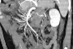
Multislice CT coronary angiography images show just far the modality has advanced since the days of single detectors and slower gantry rotation. In addition to the new hardware, researchers have optimized acquisition techniques, filling in some of the gaps that remained in spatial and temporal resolution. Software, as it has done in other modalities, has taken big steps toward making raw image data more readable.
CT angiography's tough job
There is much left to accomplish. A study just published in the October issue of the American Journal of Cardiology illustrates the typical capabilities of the modality. The researchers examined 20 heart transplant patients for vasculopathy with 64-slice CT angiography and intravascular ultrasound (IVUS).
Dr. Shawn Gregory and his colleagues from Harvard University in Boston were able to visualize 95% of the overall length of the coronary arteries without motion artifacts with 64-slice multidetector-row CT (MDCT). Compared with IVUS, 64-slice MDCT had a sensitivity of 70%, specificity of 92%, positive predictive value of 89%, and negative predictive power of 77% for detecting coronary allograft vasculopathy. In other words, pretty good considering the high heart rates typical in transplant patients, but not perfect (AJC, October 1, 2006, Vol. 98:7, pp. 877-884).
In a recent talk at the 2006 American College of Cardiology Foundation's Integrated Imaging in Clinical Cardiovascular Practice meeting in San Francisco, Dr. Mario Garcia discussed CT's future directions in heart imaging.
The Cleveland Clinic Foundation was among the first in line to install a 64-detector scanner two years ago, said Garcia, a radiologist at the foundation's Cardiovascular Imaging Center. And this year the group added a dual-source CT scanner (DSCT) (Somatom Definition, Siemens Medical Solutions, Malvern, PA).
Clinical results for dual-source coronary CT angiography (CTA) remain sparse, but a study presented this week at the American Heart Association (AHA) meeting in Chicago found that dual-source CTA eliminates most motion artifacts from CT angiography images. Thanks to 90º rotation and use of two x-ray tubes and two detector arrays, DSCT has a temporal resolution of 83 msec, yielding more motion-free images of the coronary arteries, even without the aid of beta-blockers.
The Siemens Somatom 64 can image the heart with spatial resolution of 0.625 mm and temporal resolution of 83 msec, offering "a vast improvement" over the four-slice CT machines that lurched for 45 seconds to cover the organ a decade ago, Garcia said. Because most everyone can hold still and hold their breath for eight seconds, it makes cardiac imaging easier for patients, too.
But limitations persist in CT imaging of the heart. For one thing CTA's "spatial resolution is limited to about half the rotation of the scanner," Garcia said. And the x-ray dose remains relatively hefty at 10-20 mSv per exam, though dose modulation has helped. The ability to glean functional information from CT data remains limited. And because clear acquisitions rely on the stability of multiple heartbeats, arrhythmias of any kind are a serious detriment.
"We currently require six to eight heartbeats to acquire CTA with a 64-detector scanner," Garcia said. "They have to be a very perfect six to eight heartbeats."
Even coronary artery calcium assessment -- a CT strong suit with solid clinical underpinnings -- is impaired under certain circumstances by an overestimation of calcium plaque volume, due to cardiac motion and partial-volume effect, he said.
"If the calcium overlaps more than the area of one detector, it will appear in more than one detector and appear larger than it is," and because the heart is also moving, the calcium deposit can appear larger still, Garcia said.
Certainly, there are ways to improve CT's accuracy. Garcia cited a 2004 CT angiography study in which the removal of statistical outliers boosted the results substantially.
In a study of 58 patients with suspected coronary artery disease, 16-detector CT yielded sensitivity of 77% and specificity of 97% for detecting significant stenosis. When patients with calcium scores greater than 1,000 were removed from consideration, sensitivity rose to 98% in the remaining 46 patients with low calcium scores, he said (Journal of the American College of Cardiology, September 15, 2004, Vol. 44:6, pp. 1230-1237).
The recently published Coronary Assessment by Computed Tomographic Scanning and Catheter Angiography (CATSCAN) study, led by Garcia and his team, also compared CTA to catheter angiography for stenosis detection. In all, 238 patients referred for nonemergency coronary angiography at 11 participating sites underwent 16-detector MDCT.
Sensitivity was limited by a high number of nonevaluable cases and a high false-positive rate, the authors wrote. Of 1,629 segments, 71% were evaluable by MDCT. After censoring all nonevaluable segments as positive, the sensitivity for detecting more than 50% luminal stenosis was 89%; specificity, 65%; positive predictive value, 13%; and negative predictive value, 99%.
So a negative CT is almost always useful, but a positive CT may not be true at catheter angiography, the group reported.
The results suggest that routine implementation of 16-slice MDCT in clinical practice is not yet justified, they found. As for the reported high false-positive rate, however, there were doubts about the methodology, specifically the wisdom of censoring all nonevalauble segments as positive for stenosis as part of the study methodology (Journal of the American Medical Association, July 26, 2006, Vol. 296:4, pp. 403-411).
Results are improved in 64-slice CT, which again offers an excellent negative predictive value, but the positive predictive value still lags, according to Garcia.
This month in Radiology, researchers from Switzerland found that although 64-slice CT angiography can image fast hearts accurately, minor variations in heart beat can significantly degrade image quality (Radiology, November 2006, Vol. 241:2, pp. 378-385, online publication September 11, 2006).
A recent 64-detector CTA study from the University Hospital in Aachen, Germany, showed good agreement with catheter-based coronary angiography at segment-based analysis, but only moderate agreement on a patient-based analysis, in 50 symptomatic patients with suspected coronary artery disease.
Per-patient (per-segment) analysis showed a sensitivity of 97.8% (86.7%), specificity of 50% (95.2%), positive predictive value of 93.6% (75.2%), and negative predictive value of 75% (97.7%) (European Radiology, September 29, 2006).
Today's future tech
How can spatial resolution be improved given the limits of scanner design? A new application Siemens Medical Solutions calls z-Sharp technology improves resolution by increasing the sampling rate and decreasing aliasing along the z-axis. The method yields isotropic resolution of less than 0.4 mm anywhere within the scan field and without increasing dose. Even the space between the wires in a stent could be delineated in a phantom study, Garcia said. However, he added, the study was conducted on a motionless phantom, so more motion artifact can be expected in vivo.
Dynamic volume CT envisions a flat-panel CT design in which the patient remains stationary and a wide array images an entire organ such as the heart, brain, or kidney in a single rotation.
Dynamic whole-heart perfusion contrast studies and CT angiography would be possible with the flat detectors, along with 3D-guided and augmented reality interventional procedures, Garcia said.
Flat detectors eliminate the need for collimation, and also do away with scatter radiation. However, "not many x-rays would be going through (the organ) and this will obviously limit resolution; the system will require more resolution" to compensate, Garcia said. Flat panel detectors aren't limited by coverage size; however, their temporal resolution is too low at this time to be practical for imaging a moving organ.
For now only the images' spatial resolution has been impressive, for example in the imaging of nonmoving objects such as cadavers' skulls.
How can temporal resolution be improved for cardiac imaging on today's spiral CT systems? Temporal resolution is "very closely related to spatial resolution for defining the size of features such as calcification," Garcia said. A typical motion velocity analysis shows that in late systole and peridiastole there is a period of relatively low movement in the coronary arteries.
"We reconstruct our images at diastasis, which is about 75% of the RR cycle," though there are "still significant motion artifacts if you choose diastasis," Garcia said. "In clinical practice we give beta-blockers to prolong diastasis and reduce motion artifacts. The lower the heart rate better we do. Some patients we can't get low enough."
Multicycle reconstruction can also be helpful when heart rates are very stable, and not helpful at all in the setting of atrial fibrillation, Garcia said.
A 2003 study by Manzke et al discussed the development of an adaptive scheme for the automatic patient-specific reconstruction optimization to improve the temporal resolution and image quality (Medical Physics, December 30, 2003, Vol. 30:12, pp. 3072-3080).
In 2006, Siggurdson et al detected occlusive coronary disease in heart transplant recipients with elevated resting heart rate, using multicycle reconstruction in MDCT (Journal of the American College of Cardiology, August 15, 2006, Vol. 48:4, pp. 772-778).
Dual source CT (Siemens), now commercially available, can also improve temporal resolution for imaging the heart, Garcia said. "If you have two x-ray tubes and two detectors at 90°, you only need one-quarter rotation, so you can double your temporal resolution," he said. The very promising system has yet to be clinically validated, so "time will tell" if it represents a real advance in cardiac imaging practice, he said.
The dose rises as the number of detector rows increases, so improving dose efficiency is a necessary tool for getting more image data from the radiation dose used, he said. Dose modulation systems on the market can reduce cardiac imaging doses by about half, he said.
Using techniques like dose modulation and collimation, Garcia's facility never provides more than 13 mSv for coronary CT angiography. "And when you compare that dose to perfusion imaging, it actually compares fairly well," he said. In that setting, a rest and stress nuclear perfusion study can yield as much as 30 mSv, two or three times the dose of CTA, he said.
Japanese researchers have generally had better CTA results than U.S. facilities, particularly in the Midwest where patients are much heavier, Garcia said.
One solution is prospective tube current modulation, in which "we turn our x-ray tube to 100% only during diastasis, and then lower the energy by about 20% during the rest of the cardiac cycle," he said. "The x-ray tube is flashing rather than being on all the time, and we can lower the dose by about 40% to 50%."
Thus, based on 145-mm coverage and a 15-second acquisition on a 40-detector scanner, dose modulation at 800 mAs yields the same 11.8 mSv dose as the nonmodulated scan at 500 mAs, Garcia said.
In a paper submitted to the 2005 American Heart Association meeting, Hesse et al used Doppler echocardiography to guide tube current modulation in coronary CT angiography. In 25 patients undergoing the CTA studies, a total of 296 segments were analyzed with 1.5 mm reconstruction. Sensitivity and specificity were 92% and 94%, respectively. The positive and negative predictive values were 65% and 99%, respectively, Garcia said.
Other phase selection methods include the fixed time offset, for example 500 msec before peak, or more commonly, a percentage of the RR interval is used. In 2000 Chandra, Garcia et al developed the delay algorithm, which assumes a reference heart rate of 72 bpm. As the heart rate deviates from that point, the delay is adjusted so as to identify the same physiological phase of the cardiac cycle.
Pitch can potentially be adjusted to get better data with less radiation. High pitch produces less radiation, while lower pitch produces more robust performance. To make this work there are complex technical issues to resolve, but "I think we're going to get there in two years," he said. When detector sizes are eventually increased, acquisition will be able to take place without moving the table at all, and the heart can be imaged in a single rotation with less radiation.
Plaque quantification is another promising research area, Garcia said. Recently Carrascosa et al characterized plaque morphology in 40 patients with MDCT (American Journal of Cardiology, March 1, 2006, Vol. 97:5, pp. 598-602).
In a study presented at the 2005 AHA meeting, Koyama et al generated contrast velocity maps to evaluate myocardial perfusion. Finally, multimodality co-registration, with PET/CT, SPECT/CT, and other combinations holds enormous promise for CT imaging of the heart, he said.
"In the future, CT will continue to show detector technology improvements leading to 10% to 20% reductions in dose" with improved spatial resolution, Garcia said. Lighter components will enable faster gantry rotation speeds. Also look for better tube modulation methods and myocardial perfusion assessment, he said.
By Eric Barnes
AuntMinnie.com staff writer
November 20, 2006
Related Reading
CT pioneer test-drives 256-slice scanner, August 21, 2006
Study: 16-MDCT yields too many nonevaluable artery segments, July 26, 2006
Less radiation dose exposure in cardiac CT is possible, July 6, 2006
Dual-source imaging promises better CT scanning, June 15, 2006
Prototype 256-slice CT scanner creates high-res images in a heartbeat, March 14, 2006
Motion-free heart images revealed with 256-slice CT, October 24, 2005
Copyright © 2006 AuntMinnie.com


