Dear CT Insider,
New imaging possibilities are emerging with the development of 256-slice CT -- and not only in the heart. Head and brain imaging are also getting an upgrade.
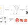
So-called area detector scanners can image the entire head and brain over 128 mm in a single rotation, enabling the fast acquisition of high-resolution perfusion images without the drawbacks of helical scanning.
Images can be acquired either as "snapshots," or sequentially in continuous acquisition mode, which can serve as a data source for whole-brain CT perfusion, subtraction CT angiography, and dynamic multiplanar reconstructions (MPRs), all in a single acquisition.
Dr. Kazuhiro Katada, chair of radiology at Fujita Health University School of Medicine in Japan, discusses his group's recent work in head and brain imaging with a work-in-progress 256-slice scanner in this issue's CT Insider Exclusive, brought to our Insider subscribers before other AuntMinnie.com members can access it.
In the realm of the small bowel, CT enterography is an exam many radiologists avoid, perhaps due to its complexity. Those willing to try it, however, are likely to find pathology that can't be seen in any other modality, according to Dr. Patrick Rogalla from Berlin. Just scroll down for the lowdown on CT enterography, along with dozens of other stories in our CT Digital Community.
By the way, Drs. Katada and Rogalla are among 64 renowned CT experts who will speak at the upcoming 9th Annual International Symposium on Multidetector-Row CT, to be held June 13-16 in San Francisco. Hope to see you there!


