From the perspective of a cardiac surgeon, the presurgical information gleaned from a CT angiography (CTA) exam of pediatric patients about to undergo surgery to repair congenital cardiac malformations is well worth the trade-off in radiation dose exposure. But just how useful is it?
To find out, Alexander Ellis, MD, a cardiac surgeon at the Children's Hospital of the King's Daughters in Norfolk, VA, completed a questionnaire rating the utility of presurgical CTA exams for each of the 33 patients he operated on at the Medical University of South Carolina (MUSC) in Charleston over the course of a year. He described the benefits of the CTA exams in the June issue of Cardiology in the Young (2010, Vol. 20:3, pp. 262-268).
The CTA exams were ordered when the surgical team felt that echocardiography images were inadequate to completely define the cardiovascular anatomy of pediatric patients. Patients ranged in age from infancy to 24 years, with a mean age of 5.6 years.
Fifteen patients had surgery to repair aortic arches, seven to repair the right ventricle to pulmonary artery conduits, seven to repair pulmonary arteries or veins, and two to repair trachea/airways. One patient needed a replacement pacemaker, and another was having a cardiac transplant.
Ellis and colleagues then analyzed the questionnaires, revealing that CTA was ranked as essential or very useful for 31 of the patients, especially with respect to presurgical planning of aortic repair for 14 of 15 patients, and pulmonary vein repair for all four patients who needed this procedure.
In addition, 14 patients were able to avoid having a cardiac catheterization, eliminating this invasive procedure and saving an estimated $7,000 to $8,000 per patient.
Information from CTA reconstructions changed the cannulation insertion site for 73% of patients who were undergoing a repeat operation and already had a right ventricle-to-pulmonary artery conduit in place. In this situation, the CTA scan helped delineate the proximity of vascular structures or conduit to the anterior bony chest wall, and may have reduced operative and cannulation complications, according to the authors.
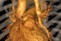
CTA-generated images also helped define or confirm complex vascular anatomy, describe anatomical relationships useful to surgical planning, and describe the severity of arterial or venous stenoses, the authors reported.
Related Reading
Pulmonary CTA finds more than pulmonary embolism in children, August 25, 2009
Radiation dose and cancer risk in pediatric CTA exams, August 21, 2009
Coronary CTA with lower tube voltage reduces radiation exposure, April 27, 2009
Low tube current, modulation reduce pediatric CTA dose, May 21, 2008
Copyright © 2010 AuntMinnie.com


