Researchers from Japan believe they have found a way to distinguish blood vessel bifurcations from solitary pulmonary nodules (SPNs) in computer-aided detection (CAD) applications for lung cancer screening.
From the viewpoint of CAD, blood vessel bifurcations and solitary pulmonary nodules look very much alike -- to the point of their generating hundreds of false-positive CAD detections in a typical lung scan.
"Since solitary pulmonary nodules are fairly common, and many of them are malignant, automated detection of SPNs is quite important for lung cancer screening," said Bin Chen from Nagoya University in Japan. "However, nodule detection methods usually cause a lot of false positives around blood vessels, especially the vessel bifurcations."
That's why the ability to distinguish vessel bifurcations and nodules is required for lung CAD systems, Chen said in a presentation at the 2010 Computer Assisted Radiology and Surgery (CARS) meeting in Geneva.
Earlier studies have focused on morphological models of nodules and bifurcations. But the enormous variability in the presentation of these vessel bifurcations means that vast numbers of models need to be built to accommodate the range of presentations, complicating development and slowing processing times, Chen said.
In addition, "nodules connected to blood vessels have a similar shape to bifurcations, so it is difficult to distinguish such nodules," he said.
Earlier this year, Fotin and colleagues developed a vessel bifurcation filter based on a third concept -- a system based on so-called "standard moments" (Proceedings of SPIE, March 9, 2010, Vol. 7624, 762413), which vastly improved the distinction of nodules compared to vessel bifurcations. But in using this method, incorrect discrimination still occurs at bifurcations with multiple side attachments in nodules with irregular shapes, Chen said. Chen proposed another path to success.
"We are proposing a bifurcation enhancement filter that is independent of nodule detection, and uses only the intensity distribution feature," he said.
The new scheme relies on object enhancement filters that are based on local intensity, a model that has gained recognition in recent years. Such filters are designed to analyze the eigenvalue distribution of a Hessian matrix for each voxel in a local region, Chen explained.
Within this enhancement framework, the research team, including Chen's Nagoya colleagues Takayuki Kitasaka, PhD, and Kensaku Mori, PhD, proposed a two-pronged solution including a line structure enhancement (LSE) filter to emphasize blood vessels and a blob structure enhancement (BSE) filter to emphasize nodules.
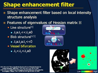 |
| A proposed enhancement filter to distinguish bifurcations from nodules is based on the eigenvalue distribution of a Hessian matrix of each voxel in a local region. The method uses both a line structure enhancement (LSE) filter and blob structure enhancement (BSE) filter to emphasize blood vessels and nodules, respectively. All images courtesy of Bin Chen. |
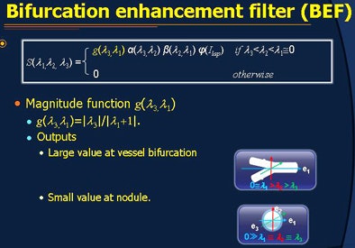
"However, since the eigenvalue distribution at the bifurcation shows such a relationship [between blood vessels and nodules], both LSE and BSE filters output ambiguous values," Chen said. To clarify the distinction of the two structures, the proposed method utilizes two likelihood functions whose output approaches 1 near bifurcations and 0 near nodules.
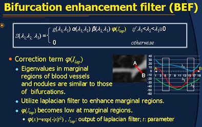
Following this calculation, a magnitude function is applied that obtains large values on bifurcations and small values on nodules, he said. Finally, because the eigenvalue distributions on the margins of blood vessels on nodules become too similar to those of bifurcation, the enhancement filter is modified to suppress enhancement in those regions.
 |
| The bifurcation enhancement filter (BEF) is based on three functions, including a likelihood function that scores bifurcations closer to 1 and nodules closer to 0. Second, a magnitude function returns large values on bifurcations and smaller values on nodules. Third, a correction function modifies the enhancement filter to suppress enhancement at areas near the margins of blood vessels, where the enhancement of nodules is similar to that of bifurcations. |
 |
 |
Chen's group applied the proposed method to 16 cases of chest CT images, containing a total of 338 nodules, both isolated and attached, ranging in size from 3.5 mm to 27 mm in diameter, Chen explained.
The noncontrast lung CT data were acquired using a slice thickness of 1.25 mm with reconstruction intervals of 2.5 mm, and the filter was applied following segmentation of the CT lung images.
Among the 200 bifurcations observed in the dataset, the bifurcation filter enhanced 183 (91%) correctly in the enhancement result. In a second experiment, the bifurcation filter was used to reduce false positives generated by a lung nodule detection algorithm.
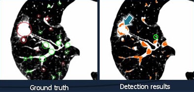 |
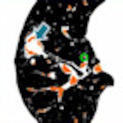
| The blood vessel bifurcation filter identified 183 (91%) of 200 "ground truth" bifurcations correctly in 16 datasets. Above, a single CT slice shows 16 bifurcations, 15 of which were detected by the BEF. Bifurcations with eigenvalues similar to those of blood vessels or nodules cannot be enhanced by the bifurcation enhancement filter. |
In this experiment, the filter reduced false positives by about 87.1% -- or from 201 per case to 26 per case, Chen said.
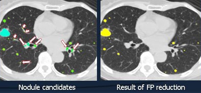 |
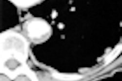
| False-positive reduction eliminated 87.1% of false positives in 16 cases. Above, a single slice of CT data shows nodule candidates at left and the results of false-positive reduction at right. |
Utilizing a novel filter that enhanced bifurcation regions, false-positive CAD detections were significantly reduced, Chen said. The experimental results show that the filter is sensitive to bifurcation and useful for reducing lung nodules.
In future work, the algorithm will be applied to automated estimation of the filter scale, and it will also be applied to the bronchus, which was excluded from the present study, Chen said.
By Eric Barnes
AuntMinnie.com staff writer
October 29, 2010
Related Reading
New CAD for COPD parses disease type, severity, October 1, 2010
Bundling of CAD software boosts lung lesion detection at CT, September 21, 2010
Combined PET/CT CAD improves lung nodule detection, August 6, 2010
Lung CAD shows promise as concurrent reader, January 30, 2009
Researchers cut false positives in lung CAD, November 10, 2008
Copyright © 2010 AuntMinnie.com


