MR enterography
Because of radiation concerns about CT enterography, European researchers began developing MR enterography in the early 2000s as an alternative for pediatric patients. The first feasibility study was published in 2005.
A single CTE exam exposes the patient to about 4 mSv of radiation, though the effective dose can be much higher. A longitudinal European study found that 15% of Crohn's disease patients accumulated more than 75 mSv from radiological procedures in 15 years. The risk of high radiation exposure doubled for patients diagnosed with Crohn's disease before the age of 17 years (Gut, November 2008, Vol. 57:11, pp. 1524-1529).
The CTE protocol served as a natural template for MRE's development, with variations added to reflect individual preferences to capitalize on MRI's inherently higher contrast resolution.
The preferred agent for bowel distention during MR enterography varies from institution to institution, according to Dr. Michael Gee, PhD, an assistant professor of radiology at Massachusetts General Hospital (MGH) in Boston. Most radiologists use a neutral enteric agent, but some Europeans prefer polyethylene glycol (PEG).
Gee's group at MGH mix a neutral enteric agent mixed with an oral suspension of ultrasmall iron oxide particles. When mixed with water in the bowel, the mixture looks dark on T2-weighted MRI. Before MRE, an intravenous gadolinium-based contrast medium is injected to enhance extraluminal tissue.
Pivotal MRE trial
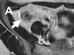
The Mayo Clinic was the site for a key efficacy trial of MR enterography. The prospective study of 23 adult patients by Dr. Hassan Siddiki and colleagues in 2009 concluded that MRE and CTE are about equally sensitive for detecting active small-bowel inflammation, though the CT's image quality was judged slightly superior (American Journal of Roentgenology, July 2009, Vol. 193:1, pp. 113-121).
Overall, CTE and MRE were both more than 90% sensitive to the presence of positive findings for Crohn's disease. CTE was somewhat more specific than MRE (88.9% versus 66.7%), but the difference was not statistically significant.
A small prospective trial by Gee and colleagues at MGH published this year found that MRE is at least as good as CTE for determining the initial presence, extent, and severity of Crohn's disease for pediatric patients (AJR, July 2011, Vol. 197:1, pp. 224-229).
The findings were based on 18 consecutive patients, all of whom were less than 18 years old. They underwent both CTE and MRE to establish an initial primary diagnosis of Crohn's disease. CTE was used as the reference standard. The sensitivity, specificity, and accuracy of MRE were 90%, 82.6%, and 86.7%, respectively.
Though MRE spares children from radiation exposure, CTE remains the first choice for many radiologists for imaging young patients. They prefer CTE because it is incredibly fast. CTE imaging can be completed in five minutes, compared with 45 minutes for an MRE evaluation. The higher speed greatly reduces the risk of motion artifacts and vomiting from children whose stomachs have been filled with oral contrast.
Diagnostic gold standard
Despite imaging advancements, histology remains the gold standard for Crohn's disease diagnosis. CTE or MRE help confirm such findings, while establishing the disease's extent and severity. After a negative biopsy in the face of persistent symptoms, enterography is used to identify inflammatory areas of the bowel for tissue sampling.
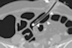
Because of superb spatial resolution, CTE is a good choice for establishing the presence and extent of active inflammatory disease. Mural stratification characterized at CTE is the most sensitive finding for active inflammatory disease, according to Dr. Khaled Elsayes, an associate professor of radiology at MD Anderson Cancer Center in Houston. Mural stratification can be appreciated by its trilaminar appearance, with enhanced outer serosal and inner mucosal layers and an interposed submucosal layer of lower attenuation.
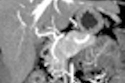
A prominent vasa recta, often referred to as a "comb sign," and increased mesenteric fat attenuation are the most specific features of active Crohn's disease described by CTE.
Though MRE also can establish these findings, its capabilities extend to water-sensitive, T2-weighted imaging that differentiates between active inflammation and fibrosis. Active inflammation on MRI is characterized by bright areas of the bowel wall on T2-weighted imaging, whereas fibrosis is characterized by a hypointense presentation and also a T2-weighted sequence.
Dynamic gadolinium-contrast enhancement also aids the assessment of bowel wall pathology, with active inflammation revealing itself from early mucosal enhancement followed by delayed enhancement of the rest of the bowel wall. Fibrosis is characterized by an absence of enhancement of the mucosa, according to Gee.
"Between the T2-weighted images and the postcontrast images, we basically get two shots to try to detect fibrosis on MRI that we wouldn't have the opportunity to look at with CT," Gee said.
The ability to differentiate between active inflammation and fibrosis is crucial for effective patient management. Active inflammation indicates the presence of reversible strictures that can be treated with anti-inflammatory drugs. Fibrotic strictures require surgical intervention to avoid future bowel obstructions, Elsayes said.
Previous page | 1 | 2 | 3 | Next page
