Tuesday, November 29 | 10:30 a.m.-10:40 a.m. | SSG05-01 | Room E353C
Researchers at the Mayo Clinic have developed a biological liver phantom for comparing perfusion imaging with contrast-enhanced CT and MR angiography (MRA).Currently, no biologically realistic model for comparing perfusion in CT and MR imaging exists, according to the researchers. The phantom has been in development since March 2010 as a multidisciplinary collaborative project between fellow graduate students in Mayo's department of radiology and biomedical engineering.
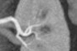
To develop the phantom, the researchers connected a fixed porcine liver to a flow-controlled perfusion circuit and submerged it in a body-shaped CT/MRI-compatible container. The phantom was perfused at various flow rates and injected with iodine contrast for CT and gadolinium contrast for MRI.
The phantom was scanned for 50 seconds using a perfusion CT protocol and an accelerated time-resolved MRA sequence. Three regions of interest from corresponding axial slices in CT and MRI were selected over the input vessel and two tissue segments.
The flow-controlled phantom experiments demonstrated consistent tissue time-enhancement curves for both CT and MRI, suggesting that this biological liver model may be useful to compare quantitative perfusion parameters between CT and MRI, according to the researchers.
"The phantom studies have given us much insight into variables that influence physiologic perfusion imaging by both CT and MRI," said Scott Thompson, a candidate in the medical scientist training program at the Mayo Medical School. "We are currently translating the knowledge gained from these ex vivo phantom studies to CT and MR perfusion experiments utilizing an animal model of liver cancer developed in our lab."


