SAN FRANCISCO - Dual-source coronary CT angiography showed high diagnostic accuracy for predicting the need for coronary intervention in what its investigators are calling the largest multicenter trial to date, according to results presented on Wednesday at the International Society for Computed Tomography (ISCT) meeting.
The group also found that image quality at dual-source CT is independent of heart rate, and despite the study's multicenter design, that the radiation dose was only moderate, said presenter Dr. Christoph Becker from Ludwig Maximilian University in Munich.
Coronary CT angiography (CTA) has been suggested to rule out coronary stenoses in patients with a low to intermediate pretest likelihood of needing a coronary intervention. However, the accuracy of dual-source CT in this patient group had not been assessed in a large-scale, multicenter trial until now, Becker said.
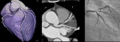 |
| A 48-year-old woman with a tight stenosis in the proximal left anterior descending coronary artery was a perfect candidate for stenting. A circumscribed lesion can be seen in the artery. Her heart rate was 58 beats per minute and she received 4 mSv of radiation. All images courtesy of Dr. Christoph Becker. |
The findings presented at ISCT come from the Multicenter Evaluation of Dual-Source CT in Patients With Intermediate Likelihood of Coronary Artery Stenoses (MEDIC) trial.
Becker, Dr. Udo Hoffmann, Dr. Suhny Abbara, and colleagues enrolled 415 patients with a 30% to 80% predilection for coronary artery disease at seven sites around the world (in Denmark, Germany, the U.S., Mexico, and India). All patients were scheduled for invasive coronary angiography and were between the ages of 30 and 80 years.
None received beta-blockers; the scans were performed using dual-source CT (Somatom Definition, Siemens Healthcare). Investigators used either 2 x 64 x 0.6-mm collimation with 0.33-sec gantry rotation or 2 x 128 x 0.6-mm collimation with 0.28-sec gantry rotation.
The protocol was based on body mass index (BMI): BMI less than 30 necessitated 100 kVp and 360 mAs, while higher BMI led to settings of 120 kVp and 360 mAs.
A core lab interpreted the results based on an 18-segment model for the prognosis of stenosis. Image quality was assessed in all the studies, Becker said. For invasive coronary angiography, there was onsite interpretation evaluated independently from CT with the option for intervention.
"Although the patients had an intermediate pretest likelihood of coronary artery disease, the majority of patients [80%] who were enrolled in the study did not receive any further coronary intervention" after being imaged with CTA, Becker said.
Another 17% of the patients went on for angioplasty or stenting with 3% of the patients receiving bypass grafting.
CTA vs. coronary intervention
|
Although the study was conducted around the world at various institutions with different protocols depending on BMI, the average radiation dose was 6 mSv, according to Becker. In addition, the image quality hovered around four, meaning the images were fully evaluable with minor artifacts.
During a discussion period at the conference, Becker noted that beta-blockers are still used at his institution despite the MEDIC results suggesting that they are not needed with dual-source CT. Beta-blockers were not used during the trial so the researchers could see the performance of CT, but they do improve image quality, he said. As such, the decision of whether to use beta-blockers is determined for each individual patient.
The MEDIC trial was sponsored by Bayer HealthCare and Siemens Healthcare, and several authors received research grants and/or speaker honorarium from Bayer and/or Siemens.


