Tuesday, November 27 | 11:20 a.m.-11:30 a.m. | SSG05-06 | Room S405AB
The use of iterative reconstruction brings important benefits, but it may have little effect on quantifying regional disease patterns in diffuse lung disease -- meaning the techniques can be used interchangeably, according to a study from University of Ulsan College of Medicine in Seoul, South Korea."Although many previous studies suggested that iterative reconstruction is useful in reducing radiation dose without increased noise, many clinical radiologists are still reluctant to use it in daily practice because the image impression is slightly different from the conventional images reconstructed with filtered back projection," explained Dr. Joon Beom Seo, PhD. "This problem is most prominent in the field of imaging of diffuse interstitial lung disease (DILD) because the diagnosis and severity assessment of DILD is based on the perception of differences in texture of local diseases."
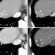
The study aimed to determine if changing the algorithm would influence the assessment of DILD at thin-section CT. The study team acquired thin-section lung CT images in 50 patients with DILD and reconstructed them with both iterative reconstruction in image space (IRIS) and filtered back projection (FBP) techniques.
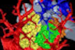
Two experienced radiologists looked at features such as ground-glass attenuation, consolidation, emphysema, reticulation, micronodules, and honeycombing using dedicated software, and fractions of disease patterns were calculated. Agreement between the techniques and between readers was high for all features and disease patterns.
"Our study showed that in quantifying local disease pattern on thin-section CT of the lung, the variation caused by changing the reconstruction algorithm was not greater than the inter- and intrareader variation," Seo told AuntMinnie.com. "This result suggests that iterative reconstruction methods can be used interchangeably with conventional FBP methods."


