"The characterization of a renal mass is a common daily problem for the abdominal radiologist," explained Dr. Achille Mileto. "Many of these lesions are serendipitously detected during imaging examinations performed for other reasons and necessitate additional testing for precise characterization."
Contrast enhancement in CT exams is the diagnostic clue that enables radiologists to distinguish between a benign nonenhancing cyst and a malignant enhancing solid mass. But contrast enhancement is an imprecise technique due to differing placement of regions of interest, and the difficulty of selecting appropriate regions of interest in intrarenal tumors, which can complicate diagnosis, he said.
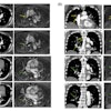
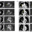
Dual-energy CT (DECT) can address some of these shortcomings because it can avoid the unenhanced image acquisition, as a single region of interest on a dual-energy color-coded iodine map shows baseline attenuation and enhancement in Hounsfield units, Mileto said.
DECT enables direct quantification of iodine concentration (in mg/mL) as a possible new paradigm for evaluating the vascularity of renal masses, rather than standard enhancement measurements. Using this technique in the study, the researchers compared calculated iodine concentrations with known iodine concentration in a phantom model and found nearly 100% correlation between the two measures.
Iodine quantification is accurate and feasible for characterizing renal masses, Mileto concluded, reducing the cost of additional tests.
