MR angiography is comparable to CT pulmonary angiography and provides good, contrast-rich visualization of pulmonary arteries to the segmental level, according to research to be presented May 12 at the International Society for Magnetic Resonance in Medicine (ISMRM) 2025 meeting in Honolulu.
The finding could eventually lead to a change in the way pulmonary embolism (PE) is diagnosed in emergency departments, especially in patients for whom CT pulmonary angiography (CTPA) is not recommended, according to a team led by Rudy Rizzo, PhD, of the University of Michigan. For their ISMRM poster presentation, the researchers investigated the detectability of PE by noncontrast MRA at 0.55 tesla.
While CTPA is considered the gold standard for rapid PE diagnosis, an alternative is needed for those who are pregnant or who cannot immediately receive iodinated contrast agents, according to Rizzo and colleagues. There has also been little exploration of noncontrast MR angiography (MRA), and research has been limited to high field strength, according to Rizzo.
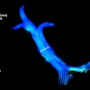
In a healthy patient diagnosed with acute PE and prescribed with anticoagulant therapy after CTPA, the group tested whether MRA could generate bright and uniform blood contrast against which blood clots appeared dark. The patient underwent the low-field MRA protocol (3D noncontrast free-breathing T2-weighted turbo spin echo sequence) 24 hours later.
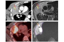
 Axial (A) and Coronal (B) CTPA show multiple partial intraluminal filling defects along the course of the right middle lobar segmental branches (medial segment) (yellow arrow) and lower lobar and postero-medial segmental branches (green arrow). Note the low-signal filling defects on the axial and coronal 0.55T MRI sequence. Caption and image courtesy of Rudy Rizzo, PhD, University of Michigan, and ISMRM
Two cardiothoracic radiologists evaluated the low-field MRI first, followed by the CTPA diagnostic reference. Using a 5-point Likert scale, both readers reported "very good overall image quality" for MRA that was comparable to CT images.
Both readers also agreed on the ability to track pulmonary arteries to the segmental level, with one reader confident up to the subsegmental level. Finally, both readers expressed high confidence in distinguishing pulmonary veins from arteries and in making the diagnosis.
While CTPA images displayed higher resolution, sharper details, and a more homogeneous white blood pool -- attributed to the imaging contrast -- MRA images showed well-opacified pulmonary arteries, though reduced resolution along the axial dimension was observed due to the large slice thickness, the team noted.
"Whole-body 0.55T MR scanners may improve MRI accessibility in high-volume settings and, with reduced B0 inhomogeneity, are promising for lung imaging," Rizzo and colleagues said, emphasizing that there is room to further explore a noncontrast MRA approach, especially in the context of the advantages of whole-body MR scanners."
Check out AuntMinnie.com’s full coverage of ISMRM 2025 here.