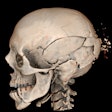
A new technique could reduce radiation exposure in children undergoing repeat CT head scans without reducing image quality, according to a study published online September 20 in the Journal of Neurosurgery.
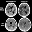
The approach works by reducing the number of slices to just seven versus the standard 32 to 40 slices per patient, reducing radiation exposure by an average of 92% while maintaining diagnostic accuracy in patients with hydrocephalus, wrote the study team from Johns Hopkins Children's Center.
The group had hypothesized that using fewer slices would also reduce clarity and diagnostic accuracy, but this turned out not to be the case. In 50 patients ages 17 years or younger, the researchers compared two standard scans with the low-dose alternative.
The radiation-minimizing technique produced clear and accurate images of the brain ventricles in all 50 patients. Still, ventricle size was affected, with the low-dose approach yielding a 4% error rate. Two of the images were visually ambiguous, and in three cases clinicians noted a change in ventricular size when there was none. However, there were no false negatives, and use of the technique would not have compromised treatment options in any case, Dr. Jonathan Pindrik and colleagues concluded.


