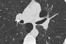
SAN FRANCISCO - Radiologists who screen smokers for lung cancer with CT should remember to look at the heart as an important predictor of mortality in its own right, according to a Tuesday talk at the International Symposium on Multidetector-Row CT (MDCT).
New studies show that mortality is strongly linked to coronary calcium, even if there are no major adverse cardiac events, and that thoracic aortic calcium can heighten the risk of stroke, said Dr. Mathias Prokop, PhD, from Radboud University in Nijmegen, the Netherlands. Considering that lung screening scans encompass the heart, radiologists must look at it and for other potential abnormalities in the image.
"One of the big issues is that we are scanning the whole chest, and the question is why should we not look at other areas? [Chronic obstructive pulmonary disease (COPD)], the heart, the bones? Lots can be done there, and you can actually use this information for many reasons" said Prokop, who heads the department of radiology and nuclear medicine in Nijmegen.
Ungated exams?
Some findings are incidental, some are important, and others are less so for getting an accurate picture of the patient's health and risk, Prokop said. For now, however, the heart is the most useful area to examine. Someday COPD will also rise in importance if reasonable treatments are developed for it.
Of course, one issue with coronary calcium is the need for gated scans to stop cardiac motion; CT lung cancer screens are ungated.
 Dr. Mathias Prokop, PhD, from Radboud University.
"But we all know that we see calcium on these [ungated] scans, and there is a good correlation between gated and ungated CT," he said. "There is a slight underestimation with ungated scans, but it doesn't mean there is always underestimation."
Some images will show more calcium than exists and some less, but generally the calcium that readers see on ungated scans is really there, Prokop said.
Even an Agatston score of zero on an image doesn't mean calcium is not present, he said. The literature shows that follow-up scans three months later often show coronary artery calcium, and the variability of the scans (about 50% for Agatston score and 36% for volume score) means that calcification was probably there from the start.
A new study in Radiology (Chiles et al, March 9, 2015) that examined the association between coronary artery calcium and mortality in the National Lung Screening Trial (NLST) found that visual assessment of lung cancer screening scans, even without Agatston scoring, was sufficient to assess cardiac risk.
In that paper, visual assessment of calcifications showed good agreement with the categorized Agatston scores (weighted κ = 0.75), and radiologists using a visual assessment assigned participants to the same risk category as the Agatston score in 73% (1,052/1,442) of CT scans and within one risk category in 99.7% of scans.
"The simplest method, an overall visual assessment of [coronary artery calcium] as none, mild, moderate, or heavy, was able to separate patients into risk categories on the basis of either [coronary disease] death or [acute myocardial infarction]," Chiles and colleagues wrote in Radiology. "All five radiologists in this study preferred the visual assessment, not only because it was faster and simpler than either of the other two methods, but it also eliminated the need for additional software."
"Visual assessment is good enough," Prokop said. "Looking just at an overall impression is good enough to give you a good estimate of the hazard of your particular patient."
Lung scans predict cardiovascular risk
But does the presence of calcification on lung screening exams predict cardiovascular risk? It does, Prokop said. Takx and colleagues just completed an examination of participants in the Nederlands-Leuvens Longkanker Screenings Onderzoek (NELSON) lung cancer screening trial (Journal of Cardiovascular Computed Tomography, January-February 2015, Vol. 9:1, pp. 50-57). They found that any calcification increases the risk of death among smokers screened for lung cancer, and that Agatston scores 400 and higher increased the risk of death by a factor of 12 -- "quite a dismal survival factor," Prokop said.
In 2012, Jacobs and colleagues found that an Agatston score of 1,000 or more on a low-dose CT scan increased the chance of dying within 2.6 years by a factor of 10, compared with a scan with no detected calcium (American Journal of Roentgenology, March 2012, Vol. 198:3, pp. 505-511).
"Coronary calcium actually measures more: It probably measures your ability to withstand other diseases, so if there's cardiac comorbidity, your chance of dying from this other disease is higher," Prokop said. And calcium is a "really massive risk predictor" when it is combined with the classic situation of hypercholesterolemia, he added. Finally, the number of calcifications is a big predictor of elevated cardiovascular risk.
Even more to look for
All-cause mortality also spikes with a rise in thoracic aortic calcification, as Buckens et al reported in European Radiology (January 2015, Vol. 25:1, pp. 132-139), Prokop said.
Buckens and colleagues showed that even though adverse coronary events are mainly determined by coronary artery calcium, if you look at noncoronary vascular events such as stroke, aortic aneurysm, and peripheral artery occlusive disease, then aortic calcium is predictive. The study also showed that osteoporosis affects survival, Prokop said.
"If you have vertebral fractures, you have twice the chance of someone who has the same other risk factors [of dying] within six years," he said.
Should calcification be reported on lung cancer screening scans? Absolutely, according to Prokop.
"Do we know that it will affect patients?" he said. "That is actually the big question mark, as there are no trials yet that show that treatment will improve survival."
That missing bit of data is especially hard to fathom considering that coronary calcium research has been around for close to 30 years, Prokop added.


