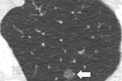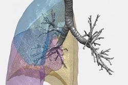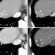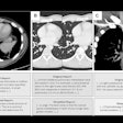
An ultralow-dose CT protocol that reduces radiation dose to 4% of standard CT can be used to evaluate some patients with chronic obstructive pulmonary disease (COPD), concludes a new study in the June edition of the American Journal of Roentgenology.
The study from Kobe University in Japan reviewed the data of 50 emphysema patients who were scanned with ultralow-dose CT using an iterative reconstruction protocol. Researchers found that the technique is effective if both iterative reconstruction and filtered back projection are used for reconstruction. The protocol could be used to stratify lung cancer risk and reduce dose for CT screening (AJR, June 2016, Vol. 206:6, pp. 1184-1192).
Concerns over excessive radiation have grown in tandem with the rising number of CT scans performed each year, and current recommendations call for using a dose as low as reasonably achievable (ALARA) while maintaining acceptable diagnostic accuracy, the authors noted. But excessive dose reduction can interfere with image analysis and interpretation.
CT is frequently performed in COPD patients, enabling the quantitative evaluation of COPD progression and helping physicians monitor therapy. Lead author Dr. Mizuho Nishio and colleagues hypothesized that ultralow-dose CT would be sufficient to evaluate most patients with the use of iterative reconstruction.
The retrospective study included 50 patients who had undergone COPD evaluation with both standard-dose CT (SDCT) and ultralow-dose CT (ULDCT) with a 320-slice scanner (Aquilion One, Toshiba Medical Systems). The tube current for standard-dose CT was 250 mA, versus 10 mA for ultralow-dose CT.
Standard-dose CT scans were processed with conventional filtered back projection, while two sets of ultralow-dose scans were processed: one set with filtered back projection and another with a commercially available iterative reconstruction protocol (adaptive iterative dose reduction [AIDR] 3D, Toshiba).
After a radiologist identified emphysema in 31 patients via visual assessment, the researchers used software they developed to quantify the extent of emphysema using three metrics: low-attenuation volume percentage, mean lung attenuation, and total lung volume.
The ultralow-dose CT technique succeeded in lowering dose dramatically, Nishio and colleagues found. The ULDCT scans had a CT dose index volume (CTDIvol) of 0.41 ± 0.095 mGy, just 4% of the CTDIvol of 10 ± 1.83 mGy for standard-dose CT.
But how well did the quantification software work with the lower-dose images? Each of the three quantification metrics produced different results, but in general the researchers found that it is probably best to use the ULDCT technique with iterative reconstruction. They also found that the AIDR 3D technique may have particular advantages not found with other iterative reconstruction techniques.
"Overall, our results validated the notion that the combined use of ULDCT without and with iterative reconstruction can yield reliable emphysema quantification and that ULDCT can substitute for SDCT for quantifying emphysema," they wrote.


