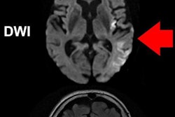
Imaging stroke patients directly in a surgical suite with conebeam CT was nearly as accurate as conventional CT and could speed up patient care by as much as an hour, according to a presentation at the annual Society of NeuroInterventional Surgery (SNIS) meeting in San Francisco.
Patients suspected of having had a stroke generally need to undergo a CT exam for diagnosis before being eligible for surgical interventions such as endovascular thrombectomy. But transferring a patient from the CT exam room to the surgical suite can delay time-sensitive treatment.
To help minimize this delay, researchers from Canada tested the ability of conebeam CT to detect hemorrhage, occlusion site, ischemic core, and at-risk tissue. They found that it compared favorably with baseline and follow-up CT scans in terms of accuracy.
"By using this [conebeam CT] technology in the angio suite, hospitals can reduce intrafacility transfer delays and hence the time of stroke symptom onset to treatment, which will significantly reduce brain damage and improve outcomes for patients," said lead author Nicole Cancelliere, an interventional clinical research technologist at Toronto Western Hospital.


