MRI scans appear to have uncovered abnormalities in certain brain pathways that may cause attention deficiencies among patients who have experienced subcortical strokes, according to a study published online May 8 in Radiology.
The keys to the discovery are the use of voxel-based lesion-symptom mapping (VLSM) and diffusion-tensor tractography (DTT), which showed an acute stroke lesion in the brain's right hemisphere. The finding correlated with prolonged reaction times among subcortical stroke patients performing an attention task.
"Impairment of attention has been observed in patients with both cortical and subcortical stroke," said senior author Dr. Chunshui Yu, from Tianjin Medical University General Hospital in China. "In cortical stroke, the direct involvement of cortical regions associated with attention may account for the deficit. However, the parts of the nervous and brain systems underlying attention deficit in subcortical stroke remain largely unknown."
Yu and colleagues enrolled 49 patients with subcortical stroke, along with 52 age-matched control participants. All subjects underwent MRI scans with a protocol that included VLSM and DTT, as well as an attention network test to evaluate visual attention function.
VLSM is designed to analyze relationships between tissue damage and behavioral deficits and also identify lesion locations related to attention deficit in stroke patients. DTT provides 3D visualization of specific white-matter tracts in the brain and can help determine impaired brain connections more than six months after a stroke.


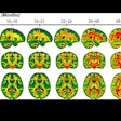
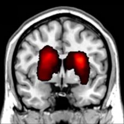
MR images provide a lesion incidence map (A) in patients with acute stroke. The map (B) also shows regions in which at least 10 patients had a lesion. Color bar denotes the probability of lesion distribution. The brain region (C) also is correlated with attention deficit in the voxel-based lesion-symptom mapping (VLSM) analysis. Color bar denotes the t-values. Images courtesy of Radiology.
VLSM results showed that the patients' delayed response time correlated with an acute stroke lesion in the right caudate nucleus and nearby white matter. DTT showed that the lesion was found in the right thalamic- and caudate-prefrontal pathways.
"The impairment of the right thalamic- and caudate-prefrontal pathways was consistently associated with attention deficit in patients with right subcortical stroke," Yu added. "Based on this association, one can estimate which patients with stroke would be more likely to develop into long-term persisting attention deficit by evaluating the lesion-induced damage to these pathways."
Thus, the findings could help determine which stroke patients would benefit from early intervention to reduce cognitive decline, Yu and colleagues noted.


.fFmgij6Hin.png?auto=compress%2Cformat&fit=crop&h=100&q=70&w=100)


.fFmgij6Hin.png?auto=compress%2Cformat&fit=crop&h=167&q=70&w=250)


