Patient-specific 3D-printed coronary models have allowed clinicians to determine ideal cardiac CT protocols for various cardiovascular diseases with greater safety and accuracy than traditional testing methods, according to a review article published online on January 24 in Current Medical Imaging Reviews.
Among the most common medical applications of 3D printing are facilitating presurgical planning and simulation, supporting medical education, and improving physician-to-patient communication. These widely explored uses of 3D printing have proved to be especially valuable in the management of cardiovascular diseases in recent years, noted Zhonghua Sun, PhD, professor of medical radiation sciences at Curtin University in Australia.
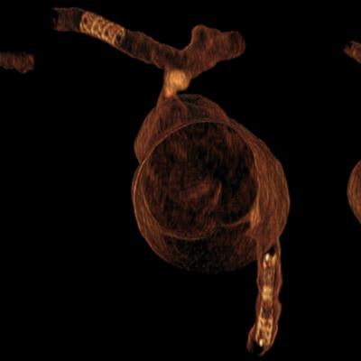
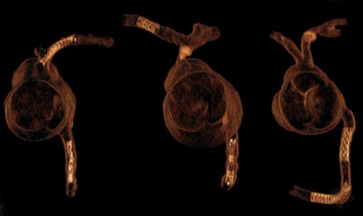 3D CT visualization of personalized 3D-printed coronary models, with coronary stents in the main left and right coronary arteries simulating coronary stenting. Image courtesy of Zhonghua Sun, PhD.
Taking a step beyond these applications, Sun investigated how researchers have been using 3D-printed heart models to optimize cardiac CT scanning protocols -- with the ultimate aim of minimizing radiation exposure to patients without lowering image quality. The review article focused on the following cardiovascular diseases:
- Coronary artery disease: Recent studies have demonstrated the potential of 3D printing to improve CT protocols for coronary artery disease. In prior research, Sun and colleagues generated patient-specific 3D-printed coronary artery models based on cardiac CT scans and fitted the models with 3D-printed calcified plaques. They subsequently scanned the 3D-printed models with different slice thicknesses to determine the best slice thickness for evaluating varying degrees of stenosis.
For patients with coronary stents, standard coronary CT angiography offers restricted visualization due to beam hardening artifacts. This limitation has led clinicians to turn to high-resolution CT scanning with postprocessing image reconstruction. A recent study by Sun and colleagues demonstrated the value of using 3D-printed coronary models with stents to compare different scanning and reconstruction protocols in search of the ideal one for imaging coronary stents.
- Aortic disease: Diagnostic assessment of aortic diseases such as aortic dissection depends heavily on CT angiography. Burris et al recently showed that measuring changes to aneurysms in patients with aortic dissection, especially in those who underwent endovascular repair, can be more accurate on 3D-printed models of the patient's aorta than on their standard CT scans.
- Pulmonary embolism: In a previously published report, Sun and colleagues acquired multiple rounds of CT pulmonary angiography (CTPA) scans of 3D-printed pulmonary artery models that contained animal blood clots. CTPA is considered a first-line technique in diagnosing pulmonary artery disease. The researchers used a wide range of peak kilovoltage (kVp) and pitch values for the scanning rounds. This process enabled them to identify an ideal set of kVp and pitch parameters for diagnosing pulmonary embolism -- one that used 80% less CTPA radiation dose than the standard protocol without negatively affecting image quality.
"These early results showed that 3D-printed models can serve as a useful tool for testing different CT scanning techniques ... [and] are promising for achieving the significant reduction of radiation dose while maintaining diagnostic image quality," Sun wrote.
3D printing offers researchers a reliable alternative for optimizing CT scanning techniques that may be more cost-effective than purchasing commercial anatomical phantoms and safer than performing scans on human subjects, he concluded.


