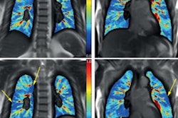
Particular CT imaging findings such as a diffuse alveolar damage (DAD) pattern can help radiologists diagnose electronic cigarette or vaping product use-associated lung injury (EVALI), according to a presentation delivered on Monday at the virtual RSNA 2020 meeting.
By flagging these findings, radiologists can alert their clinician colleagues that an EVALI diagnosis should be considered, presenter Dr. Timothy Mickus of Allegheny Health Network in Pittsburgh told session attendees.
"EVALI patients often present with nonspecific clinical symptoms that mimic viral illness but rapidly progress," he said. "Respiratory complaints are common, as are gastrointestinal problems, fever, and fatigue."
Electronic nicotine delivery systems were introduced into European and U.S. markets between 2006 and 2007. Their use is most common among young people: In fact, a 2018 survey conducted by the National Youth Tobacco Survey found that 20.8% of high school students have tried or were currently using e-cigarettes. In 2019, there was an EVALI outbreak that prompted the U.S. Centers for Disease Control and Prevention to declare it an epidemic among high school patients.
"Instead of burning tobacco leaves, these devices aerosolize or vaporize nicotine-containing liquids, often with added flavorings called juices," Mickus said. "They're also being increasingly used to aerosolize marijuana and [tetrahydrocannabinol (THC)] products."
General clinical criteria for diagnosing EVALI include a history of vaping within 90 days, abnormal findings on chest CT, and exclusion of other potential disease causes like infection, autoimmune disease, or malignancy, he said. But there are particular imaging findings that radiologists can identify and share with colleagues to help come to a more definitive diagnosis of EVALI, including the following:
- Diffuse alveolar damage/pneumonia. Common imaging features include multifocal/diffuse ground-glass opacity and/or consolidation; subpleural sparing, both peripheral and central; and septal thickening.
- Subacute hypersensitivity pneumonitis pattern. This occurs in about a third of cases, according to Mickus. Imaging features include ground-glass opacity, poorly defined cetrilobular nodules, and mosaic attenuation/air-trapping/lobular low attenuation.
- Acute eosinophilic pneumonia. This is often seen as the result of binge-smoking traditional cigarettes, but it also manifests through e-cigarette usage, Mickus said. Imaging features include bilateral and symmetric ground-glass opacities and/or consolidation, prominent septal thickening, and pleural effusions.
- Diffuse alveolar hemorrhage. Imaging features are often asymmetric and include consolidation and ground-glass opacity, "fluffy" centrilobular nodules, and septal thickening.
"E-cigarette use is very common -- probably more common than we expect, especially in younger patients," Mickus concluded. "Clinical presentation can be difficult and nonspecific, but imaging patterns can be helpful, especially if we see a DAD pattern with subplerural sparing."


