Automated multiorgan CT analysis identifies those individuals who are at high risk of type 2 diabetes and other cardiometabolic comorbidities, researchers have found.
A team led by Yoosoo Chang, MD, PhD, of Sungkyunkwan University School of Medicine in Seoul, Korea, found in particular that the index of visceral fat showed the highest predictive performance for diabetes, outperforming traditional risk factors. The results were published August 6 in Radiology.
"The results are encouraging as they demonstrate the potential of expanding the role of CT imaging from conventional disease diagnosis to opportunistic proactive screening," study senior author Seungho Ryu, MD, PhD, also of Sungkyunkwan University School of Medicine, said in a statement released by the RSNA.
It's understood that CT imaging conducted for other indications shows promise for predicting cardiometabolic disease risk, but its ability to do so needs more exploration, the team wrote.
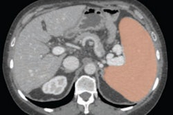
To this end, Chang's group assessed the ability of automated CT-derived markers to predict diabetes and associated cardiometabolic comorbidities via a study that included 32,166 Korean adults who underwent PET/CT exams between January 2012 and December 2015. The team tracked CT markers such as visceral and subcutaneous fat, muscle, bone density, liver fat, and aortic calcification and evaluated their predictive ability using the area under the receiver operating characteristic curve (AUC) and the Harrell C-index. The incidence of diabetes among the study cohort was 6% at baseline and 9% at median follow-up of seven years.
The group found that patients' visceral fat index had the highest predictive value for prevalent and incident diabetes, and that combining visceral fat, muscle area, liver fat fraction, and aortic calcification improved predictive performance.
| Performance of CT markers for predicting diabetes, cardiometabolic comorbidities | ||
|---|---|---|
| Marker | AUC | C-index (with 1 as reference) |
| Visceral fat index for identifying diabetes | ||
| Women | 0.82 | 0.82 |
| Men | 0.7 | 0.68 |
| Combination of visceral fat, muscle area, liver fat fraction, and aortic calcification | ||
| Women | -- | 0.83 |
| Men | -- | 0.69 |
| Visceral fat index for identifying metabolic syndrome | ||
| Women | 0.9 | -- |
| Men | 0.81 | -- |
The group also reported that CT-derived markers identified ultrasound-diagnosed fatty liver, coronary artery calcium scores greater than 100, sarcopenia, and osteoporosis, with AUCs ranging from 0.8 to 0.95.
The study findings highlight the potential of CT-derived markers to improve diabetes screening and risk assessment, according to Ryu.
"By integrating these advanced imaging techniques into opportunistic health screenings, clinicians can identify individuals at high risk for diabetes and its complications more accurately and earlier than the current approach," he said. "This could lead to more personalized and timely interventions, ultimately improving patient outcomes."
Chang's and colleagues' work adds to "the growing body of evidence in support of opportunistic CT screening," wrote Perry Pickhardt, MD, of the University of Wisconsin in Madison, in an accompanying commentary.
"In addition to addressing important research questions, the ultimate clinical implementation of these tools should add substantial value by simply leveraging data already embedded within all routine CT scans," he noted.
The complete study can be found here.


