Opportunistic chest CT could be a useful alternative to dual-energy x-ray absorptiometry (DEXA) for assessing low bone mineral density among women, the elderly, and lower-weight patients, researchers have reported.
Targeting the correct population for use of CT for this indication is key to better patient care, wrote a team led by Jiongfeng Zhang, MD, of Nanchang University in Jiangxi, China. The findings were published on May 11 in Clinical Interventions in Aging.
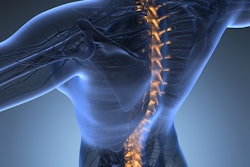
"CT scans can identify vertebral compression fractures from reconstructed sagittal images, providing an opportunity for detection and proper management," the group explained.
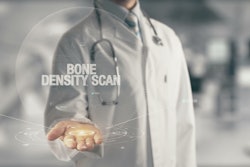
By 2050, the incidence of bone fractures in China will reach almost 6 million cases, translating to more than $20 billion U.S. dollars, the group noted. The most common imaging exam used to diagnose osteoporosis is lumbar and hip DEXA, yet more than 80% of individuals with bone fractures don't undergo bone mineral density testing, and DEXA's effectiveness can be limited, the authors explained.
That's why the opportunistic use of chest CT to assess bone mineral density via the attenuation value of Hounsfield unit (HU) expression in trabecular vertebrae shows promise, the investigators wrote.
"Chest CT scan is widely used in the screening of lung cancer and follow-up of pulmonary nodules, and its images carry valuable potential information about vertebral quality, especially sagittal images, which play an important role in the evaluation of vertebral morphology," the team explained.
Which patients would most benefit from this use of CT? Zhang and colleagues investigated the question via a study that included 1,268 patients who underwent both chest CT and DEXA within a one-month time frame between November 2020 and August 2021. The team measured CT attenuation values of trabecular bone in the midsagittal plane from thoracic vertebra 7 and used the area under the receiver operating characteristic curve (AUC) measure to assess the exam's performance for this indication.
It found the following:
- The AUC for diagnosing low bone mineral density was larger in women than in men, at 0.894 versus 0.744 (p < 0.05).
- The AUC increased gradually as individuals aged but decreased with increases in height and weight (p < 0.05).
- In women, when the researchers adjusted specificity to 90%, a threshold of 140.25 HU showed a sensitivity of 69.3%, higher than the sensitivity of 36.5% in men for distinguishing low-bone mineral density from normal.
- At age 70 or above when specificity was adjusted to 90%, a threshold of 126.31 HU showed a sensitivity of 76.1%, which was higher than that of other age groups.
The bottom line is that "the use of opportunistic CT as a screening tool identifying undiagnosed patients at high risk of fractures is without any doubt a tool that should be increasingly implemented in common practice, and may also replace the DEXA [if] the latter is not feasible," the researchers concluded.
The complete study can be found here.