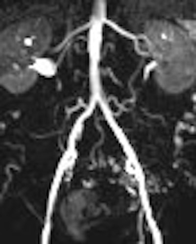
Using a parallel acquisition technique can reduce the amount of gadolinium needed for intra-arterial MRI, according to Swiss imaging experts. Furthermore, a high contrast-to-noise ratio (CNR) can be achieved in the vessel lumen, they wrote in the American Journal of Roentgenology.
"Blood gadolinium concentration must meet a target range during MR angiography data acquisition to achieve adequate arterial enhancement.... Because of the short half-life of gadolinium chelates in the circulating blood, several injections are mandatory during endovascular interventions," explained Dr. Silke Potthast and colleagues from the University Hospital Basel and the University of Basel (AJR, March 2007, Vol. 188:3, pp. 823-829).
Potthast's group proposed a faster acquisition technique for reducing the amount of gadolinium. In their study, they evaluated nine patients with 3D FLASH MR angiography (MRA) and the alternative technique, generalized autocalibrating partially parallel acquisition (GRAPPA). They also compared the two MR techniques in an aortic pulsatile flow phantom.
Of the nine patients, the majority had arterial occlusive disease of the iliac axis. The patients underwent angiography and angioplasty with a 6-French introducer left in the common femoral artery.
For the FLASH protocol, the acquisition time was 20.3 seconds using 81.2 mL of gadoterate meglumine (Dotarem, Guerbet, Roissy, France). For the GRAPPA protocol, the acquisition time was 14 seconds with 56 mL of contrast agent.
For the patients, maximum intensity projections (MIPs) were calculated from the contrast-enhanced images. Image quality for evaluating the renal arteries was graded on four-point scale (1 = excellent, 4 = poor). In the phantom, intraluminal signal intensity was measured distal to the tip of the catheter along the tube.
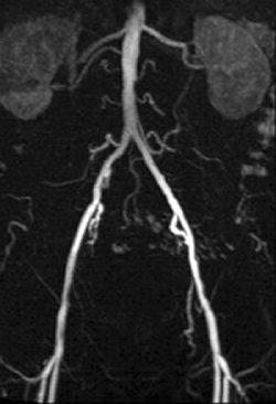 |
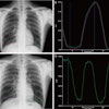
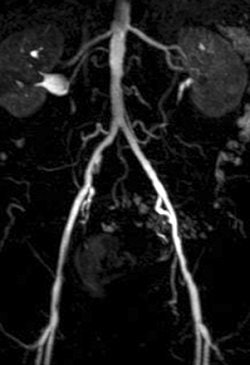
| A 78-year-old man with peripheral arterial occlusive disease. Lumbar arteries are evident with both MRI acquisition techniques. Iliac axis has good runoff on both sides after percutaneous transluminal angioplasty (above). Anteroposterior maximum intensity projection 3D fast low-angle shot (FLASH) intra-arterial MR aortogram obtained with standard technique (below). |
 |
| Potthast S, Bongartz G, Huegli R, Schulte A, Schwarz J, Aschwanden M, and Bilecen D. "Intraarterial Contrast-Enhanced MR Aortography With and Without Parallel Acquisition Technique in Patients with Peripheral Arterial Occlusive Disease" (AJR 2007; 188:823-829). |
According to the results of the patient study, both techniques were well tolerated and had no side effects. In addition, the diagnostic value of GRAPPA MRA was given an average grade of 2.2 compared to 2.0 for FLASH MRA. Using the parallel acquisition technique, the CNR ranged from 32.0 in the renal arteries to 125.5 in the aorta.
In the phantom, the mean CNR ranged from 290-717 with GRAPPA MRA and 230-585 with FLASH MRA. A 15% to 26% increase in CNR was calculated for the GRAPPA MRA scans.
"Adequate enhancement of signal intensity in the arterial lumen is mandatory for the diagnosis of stenosis and for catheter tracking with the help of road maps," the authors wrote. "In theory, parallel acquisition technique is used at the cost of CNR.... (In this study) higher CNR was obtained for parallel acquisition MR angiography in the aortic flow phantom."
Overall, the 30% reduction in acquisition time led to a 30% reduction in contrast agent consumption, they noted. Using this parallel technique could also produce cost savings, the authors added.
By Shalmali Pal
AuntMinnie.com staff writer
March 21, 2007
Related Reading
1.5T, 3.0T MRI are equivalent for assessing myocardial infarction, March 15, 2007
Dual-contrast MRI detects and shows extent of DVT, March 14, 2007
Scottish study shows potential gadolinium-NSF link, March 9, 2007
Copyright © 2007 AuntMinnie.com

