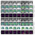
More than 100 intensive care patients were included in this German study. Patients received bedside chest radiographs using four acquisition techniques: CR and DR with and without a grid. Image quality was then evaluated by four radiologists using a nine-point visibility scale. They evaluated lung parenchyma, soft tissues, thoracic spine, foreign bodies, and overall image quality.
Image quality of the DR images with a grid was significantly higher than quality without a grid for all structures, the researchers found. The use of a grid in CR significantly improved overall image quality and lung parenchyma and soft-tissue delineation, but foreign body delineation was better without a grid.
The visibility scores for DR images were significantly higher than the CR scores for all structures.
"We are very interested in improving the diagnostics in intensive care medicine, [and] we have now improved the technological basis especially for the flat-panel technology," Dr. Thomas Vogl, PhD, from the University of Frankfurt, told AuntMinnie.com.
The system also already has high clinical acceptance, he added.


