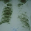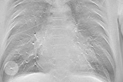
Radiologists who interpreted images acquired with a digital radiography (DR) tomosynthesis system were nearly four times more likely to find chest nodules than those interpreting conventional DR studies -- and tomo's edge grew as the nodules got smaller, according to a new study in Radiology.
Lead author James Dobbins, PhD, from Duke University and researchers from other centers compared DR with tomosynthesis to DR without tomo in a group of 158 individuals, 115 of whom had CT-confirmed nodules and 43 of whom were nodule-free. Radiologists using tomo had nearly four times the sensitivity for all nodules, as well as nearly eight times the sensitivity for nodules 4 mm to 6 mm -- which are difficult to determine how to manage (Radiology, July 19, 2016).
 James Dobbins, PhD, from Duke University.
Dobbins et al believe the findings are encouraging in that the study was designed to approximate how tomosynthesis would be used in a clinical environment. They also hinted that tomo could be useful for indications such as following up suspicious nodules detected on CT lung cancer screening exams.
Clinical potential outside the breast
Already a hot technology for breast imaging, previous studies have indicated that tomosynthesis could also have applications in areas such as general chest imaging by enabling radiologists to see around structures like bones that might obstruct pathology. For example, research studies have found that tomo can triple the sensitivity of radiologists using it to detect pulmonary nodules, compared with the use of conventional chest x-ray, the authors noted.
Before tomo arrived on the scene in 2006, radiologists experimented with another advanced x-ray technology, dual-energy (DE) imaging, as a way to improve visualization by taking advantage of the different energy absorption properties of bone and soft tissue. Such studies typically involved performing two x-ray exams on patients at different energy levels, with bone subtracted from the resulting studies.
The team led by Dobbins therefore sought to compare tomosynthesis with conventional DR chest x-ray, both with and without dual-energy imaging, using a commercially available tomo unit (VolumeRad, Discovery XR656, GE Healthcare). In particular, they wanted to find out how tomo might affect the management of individuals with suspicious lung nodules, and also if tomo's performance varied based on different nodule characteristics such as size.
The researchers collected data from studies performed on people at four tertiary care sites, three in the U.S. and one in Sweden. Study subjects had been referred for or had already received chest CT scans as part of their standard workup for either suspicious pulmonary nodules or clinical indications unrelated to pulmonary nodules. In all, 158 individuals were included in the study; 115 had pulmonary nodules confirmed with CT, and 43 turned out to be nodule-free.
After CT images were acquired for clinical workup, individuals underwent imaging for the research study, including conventional posteroanterior (PA) and lateral chest radiographs, dual-energy radiographs emphasizing both bone and soft tissue, and digital tomosynthesis images.
For the DE acquisitions, low-energy images were acquired at 60 kVp and the high-energy images were acquired at 120 kVp. Tomo images were acquired at 120 kVp with the individual in the PA orientation, with 60 low-exposure projections acquired in a 30° arc of the x-ray tube. Subjects then received conventional DR radiographs using each institution's standard protocol, typically 110 kVp to 130 kVp.
Images were interpreted by five radiologists, who were selected based on their different areas of specialty training and the amount of time they spent reading chest images in their daily practice, ranging from 10% to 100%. Only one reader had experience in reading dual-energy images, and none of the five had previous experience with tomosynthesis. The idea was to approximate the typical radiology environment, where not every reader is an expert in chest imaging, Dobbins told AuntMinnie.com.
In analyzing the results, the researchers chose to parse the data in three ways to measure tomo's effect:
- Nodule detection on a per-nodule basis
- Nodule detection on a per-person basis
- Overall case management
With respect to per-nodule performance, the researchers compared tomo with and without dual energy to conventional DR with and without dual energy. Two of the main metrics they used were lesion localization fraction (LLF), roughly analogous to sensitivity, and nonlesion localization fraction (NLF), comparable to false-positive rate. With LLF, higher numbers indicate higher sensitivity, while higher NLF numbers indicate higher specificity, as indicated in the table below.
| Tomo vs. conventional x-ray for all nodule sizes, 3-20 mm | ||||
| Metric | Conventional chest x-ray | Conventional chest x-ray with dual energy | Tomo | Tomo with dual energy |
| Maximum LLF | 0.038 | 0.057 | 0.135 | 0.134 |
| Maximum NLF | 0.260 | 0.367 | 0.256 | 0.265 |
The figures indicate that tomosynthesis without dual energy had a maximum LLF (sensitivity) that was 3.6 times higher than that of conventional x-ray without dual energy -- similar to previous research, according to Dobbins. Meanwhile, the maximum NLF (specificity) of tomosynthesis without dual energy was comparable to that of conventional chest x-ray.
With respect to dual-energy imaging, there was no statistically significant difference in the figures for tomosynthesis or conventional chest x-ray regardless of whether the technology was used.
The researchers found even more dramatic results when analyzing the data by nodule characteristics, particularly nodule size. For example, for 4- to 6-mm nodules, readers using tomosynthesis had 7.6 times the maximum LLF compared to conventional chest x-ray (0.017 for conventional x-ray versus 0.129 for tomosynthesis).
Similar findings were discovered with the per-case analysis, although they were not quite as dramatic as the lesion-level analysis. For all nodules 3 mm to 20 mm in diameter, tomosynthesis showed a 49% improvement over conventional x-ray (maximum LLF of 0.412 for conventional x-ray versus 0.614 for tomo).
What does it all mean?
The study is significant in that it represents the culmination of the Duke group's work on tomosynthesis, Dobbins said. Previous research had focused on the physics and technical parameters of tomo, while this paper is more oriented toward clinical use, using a new tomo data reconstruction algorithm that GE recently developed for commercial application.
Dobbins is particularly intrigued by tomo's superiority for lesions 4 mm to 6 mm. This is the lesion size at the boundary of criteria specified by Fleischner Society guidelines for following up suspicious lesions with additional testing.
"That means that tomo is very well-tuned to improving performance right at that threshold level where you are going to need to decide whether a patient has to come back for follow-up imaging," Dobbins said.
The results also hint that tomosynthesis could have a role in CT lung cancer screening, following up on suspicious lesions detected on CT screening but at a much lower cost and radiation dose.
"Much more work needs to be done before we can say definitely that tomo would be good for lung cancer screening or for this kind of follow up in place of CT, but this study suggests that tomo could be well-tuned for this application," he said.
Study disclosures
The researchers received funding from GE for the study.


