Thursday, December 5 | 11:40 a.m.-11:50 a.m. | SSQ11-08 | Room N229
Clinical DICOM headers contain a wealth of information that imaging centers can use to optimize image quality and improve digital radiography equipment utilization.Those perspectives are just a few of the messages set to come from Zaiyang Long, PhD, an assistant professor of medical physics at the Mayo Clinic in Rochester, MN, in this Thursday talk.
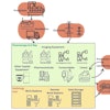
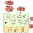
Long and colleagues created an architecture and tools to receive and extract DICOM header information from clinical digital radiography (DR) images. More specifically, the researchers took data from 18 DR systems, including study and image times, software information, voltage measurements (kV), exposure mode, and exposure time index, as well as processing-related information such as processing name, contrast and edge enhancement, and even the equipment serial numbers.
The ultimate goal was to use the information to standardize daily practices and processes and improve equipment utilization.
In one case, the researchers found grids were not used as expected for several exams with a gridded technique. As another example, many exam views showed variation in image processing selection and identified where system defaults were not currently set or where technologists were struggling to consistently achieve good image quality with default processing.
The clinical DICOM headers provided us with "valuable information on equipment usage, image acquisition, and processing," Long and colleagues concluded. "These data have been successfully used for quality improvement, technologist education, and equipment purchase decisions."
