Can chest x-rays be used to detect unsuspected coronary artery diseases? An artificial intelligence (AI) program has shown it's more than possible, according to research published June 17 in Radiology: Cardiothoracic Imaging.
In a proof-of-concept study, radiologists at Johns Hopkins University in Baltimore developed a deep convolutional neural network (DCNN) that not only identified coronary artery calcium on chest x-rays but also correlated the findings with a higher risk of cardiovascular disease. What's more, it worked independent of traditional risk factors, such as smoking or diabetes.
"We demonstrate that DCNNs can predict the likelihood of [coronary artery calcium] on chest radiographs, albeit with modest performance, and that this correlates with patient-specific cardiovascular risk," wrote the authors, led by Dr. Peter Kamel.
Machine learning has previously been used in the automated calculation of Agatston scores from electrocardiographically gated and nongated cardiac CT scans. However, CT scans have higher cost and radiation exposure than more commonly obtained chest x-rays. An automated method to screen for coronary artery calcium on chest x-rays could provide clinically meaningful prognostic information in patients with unsuspected coronary artery disease, according to the researchers.
In this study, Kamel and colleagues asked whether an automated model could be developed to predict the presence of coronary artery calcium on chest x-rays and correlate the results with cardiovascular risk.
They obtained imaging data from 1,010 patients who had undergone both standard calcium score CT scans and lateral chest x-rays within a 12-month period between 2013 and 2018. The CT calcium scoring scans were for purposes of cardiovascular risk stratification, while chest x-rays were acquired for various indications, such as chest pain, cough, and shortness of breath.
Total Agatston calcium CT scores and the scores for each coronary artery (left main, left anterior descending, left circumflex, and right coronary arteries) were determined to establish ground truth and used to train, validate, and test the program on x-rays.
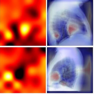
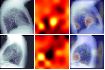 Lateral chest radiographs labeled with total Agatston scores. Despite training only on calcium score numbers, algorithms learned to focus on and prioritize the cardiac silhouette to predict the presence of coronary artery calcium. Left column, radiographs; middle column, attention-based heat maps; right column, heat maps overlaid on radiographs. Image courtesy of
 Radiology: Cardiothoracic Imaging.
The dataset included 888 x-ray images where the corresponding CT had a nonzero calcium score and 382 images with a calcium score greater than 100. The performance of the AI model for classifying between 0 and nonzero total calcium scores on the x-rays reached an AUC of 0.73 on frontal radiographs, with similar performance on laterals (AUC, 0.70). The model achieved an AUC of 0.73 classifying between zero and nonzero Agatston scores on posteroanterior chest x-rays.
| AUCs for AI classifications between 0 and nonzero total calcium scores on x-rays | |||||
| Total score | Left main | Left anterior descending | Left circumflex | Right coronary artery | |
| Posteroanterior chest x‑ray | 0.73 | 0.66 | 0.62 | 0.66 | 0.71 |
| Lateral chest x‑ray | 0.70 | 0.64 | 0.62 | 0.75 | 0.69 |
| p-value | 0.56 | 0.84 | 1.0 | 0.15 | 0.75 |
The results are modest, according to the researchers. However, in addition, frontal radiographs that tested positive for coronary artery calcium correlated with a higher 10-year atherosclerotic cardiovascular disease risk of 17.2%, compared with 11.9% for a negative test, they found.
"An automated method to screen for [coronary artery calcium] on chest radiographs may provide clinically meaningful prognostic information in patients with unsuspected coronary artery disease," Kamel and colleagues wrote.
For instance, chest x-rays are routinely obtained in the initial evaluation of chest pain in the emergency department and an algorithm that predicts the presence of coronary artery calcium could be useful in guiding subsequent diagnostic evaluation in an acute setting, they suggested.
The researchers noted several limitations. Upon review of heat maps, cardiac defibrillators, which were seen in 61 images (4% of the dataset), were confounders and sometimes more important for the algorithm's holistic decision-making than visualization of calcium.
Nonetheless, the study serves as a proof-of-concept that DCNNs can predict coronary artery calcium and cardiovascular risk on chest x-rays beyond typical human radiologist attention, the researchers noted. The results of this study may serve as a pilot for future applications in deep learning.
"Here we demonstrate the ability to extract information from medical imaging not readily noted on human review that may affect clinical management and diagnostic workup," they concluded.

