Are uncooperative children receiving more radiation exposure during digital chest x-ray? Not intentionally, of course, but imaging children in supine positions to constrain motion and reduce image distortions results in higher doses than when patients are standing, according to a study published August 5 in PLOS One.
Chinese researchers analyzed radiation doses children received in three different body positions during diagnostic digital x-ray exams: supine anterior-posterior (AP) projection, standing AP projection, and standing posterior-anterior (PA) projection. They found children in the supine anterior-posterior position received higher doses, while those in the standing posterior-anterior position received the lowest dose.
"The radiation dose to superficial organs may be lower with standing [PA projection] than with standing [AP projection] during chest [digital radiography]. Standing [PA] should be selected for chest [digital radiography] in 3- and 4-year-old children, as it may decrease the required radiation dose," wrote radiologist Dr. Bi Liu of Children's Hospital of Chongqing Medical University and colleagues.
Digital chest x-ray is used to diagnose respiratory illness in children, but studies suggest that because they are more sensitive, radiation-induced cancer may be up to three times higher in children than adults. Standing anterior-posterior and standing posterior-anterior digital chest x-rays are primarily used for cooperative children, while the supine anterior-posterior projection is often selected for uncooperative children because it constrains their motion and results in better image quality, the authors wrote.
Previous studies have looked at various combinations of tube voltage and current to optimize radiation exposure during digital chest x-rays, yet few have looked at whether the type of projection used influences the radiation dose in children, according to the authors.
In this prospective study, the researchers included 240 children (age range, 3-4 years) referred to their hospital for digital x-ray exams between July and December 2019. The children were divided into three equal groups: children who exhibited good cooperation were divided into a supine AP group, a standing AP group, and a standing PA group. Participants who exhibited poor cooperation were placed in the supine AP group.
The irradiation field was minimized as much as possible and included the apexes of the lungs, clavicles, and the costophrenic angle and the edges of the thoracic walls. The dose area product (DAP) and the entrance surface dose (ESD) were recorded after each exposure.
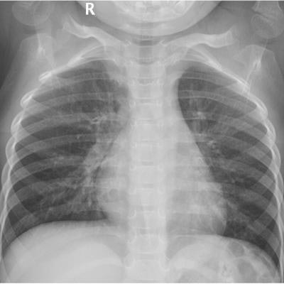
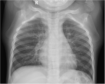
 Digital chest x-ray of a pediatric participant in the supine anterior-posterior projection group. The image shown is that of a 38-month-old female with a height of 109 cm, a weight of 15 kg, and a body mass index (BMI) of 12.63 kg/m2. The ESD value was 0.07 mGy and the DAP value was 0.22 dGy*cm2.
The researchers found statistically significant differences in DAP and ESD among the three groups. The DAP and ESD values for the standing PA and standing AP groups were significantly lower than those for the supine AP group.
| Digital x-ray radiation dose for supine AP, standing AP, and standing PA positions | |||
| Supine AP | Standing AP | Standing PA | |
| Entrance surface dose | 0.16 mGy | 0.12 mGy | 0.12 mGy |
| Dose-area product | 0.55 (dGy*cm2) | 0.42 (dGy*cm2) | 0.41 (dGy*cm2) |
"These results suggest that the use of standing [PA] can decrease the radiation dose and fully display the lung field in 3- and 4-year-old children," the authors wrote.
The pulmonary field area was also significantly higher for the standing PA group than for the standing and supine AP groups, the researchers added.
"When choosing an imaging projection for [digital chest x-ray] for 3- and 4-year-old children, diagnostic requirements should not be the only consideration; the individual's status should also be considered. Standing [PA] should be the preferred projection choice, followed by standing [PA] and supine [AP], because standing [PA] can decrease the radiation dose received during chest [digital x-ray]," the authors concluded.


