Researchers in Ethiopia have developed a new strategy for diagnosing tuberculosis (TB) in low-resource, high-incidence settings: Photographing film x-rays to create digital files and then feeding them to AI.
In a pilot study that included 498 such images, radiologists detected up to 99 cases indicative of TB, while an AI model detected 81 cases, noted lead authors Zerubabel Desita, MD, and Temesgen Tadesse, MD, of the University of Gondar, and colleagues.
“Successful mobile phone-based analog radiographic image analysis could represent an easily implementable tool for screening and reduce diagnostic barriers for people living in low-resource settings,” the group wrote. The study was published January 21 in Mayo Clinical Proceedings: Digital Health.
With an estimated 10.8 million people experiencing TB around the world in 2023 and 1.09 million deaths that year due to the disease, TB remains a major threat to global health. Previous studies have shown that AI models can improve the detection and diagnosis of TB on chest x-rays in areas where experienced radiologists are scarce, the authors explained. Yet these studies have focused on the analysis of digital images, which limits the technology’s applicability in settings where chest x-rays are available only in analog form, the researchers noted.
The group aimed to assess the diagnostic value of an AI model (qXR, Qure.ai) for TB using film x-rays photographed by mobile phones or digital cameras. The analysis used 498 images from patients seeking help for TB symptoms between January 2017 and March 2018 at health centers in Ethiopia and Guinea-Bissau who had a final diagnosis by clinical or laboratory tests.
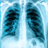
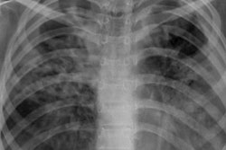
 A sample input analyzed by the AI model. The film chest x-ray was photographed with a Sony Cyber-shot 20.1 MP digital camera.Mayo Clinical Proceedings: Digital Health
Finally, the agreement regarding TB-related findings between the radiologists combined (kappa = 0.45) and each radiologist and the software (kappa = 0.56) was moderate, the researchers reported.
“Our study revealed that [the AI model] performs comparably to experienced radiologists when it is applied to [chest x-ray] films, photographed by mobile phones and a digital camera with similar sensor resolutions,” the group wrote.
At present, due to a lack of resources in low-income countries in Africa and elsewhere, it appears unlikely that the World Health Organization (WHO) will reach its goal of reducing the incidence of TB by 90% by 2035, the researchers noted.
Thus, while this was a pilot study that requires further validation, it highlights the potential use of AI to lessen the detection gap, they wrote.
“AI-guided [chest x-ray] interpretation via mobile phones may guide health care workers with no or limited training in assessing [chest x-rays] for possible TB,” the group concluded.
The full study is available here.