Researchers have trained an artificial intelligence (AI) algorithm to estimate sex on digital knee x-rays using a skeleton collection belonging to the Smithsonian's National Museum of Natural History in Washington, DC.
A team led by Dr. Petteri Oura, PhD, a forensic medicine specialist at the University of Helsinki in Finland, said the algorithm estimated sex correctly in more than 90% cases from a skeleton collection at the museum. The proof-of-concept study establishes that AI could help aid in forensic analysis of skeletal remains, they wrote.
"Combining radiographic data with automated and externally validated algorithms may establish valuable tools to be utilized in forensic anthropology," the group noted in a study published February 1 in Legal Medicine.
When analyzing unknown skeletal remains, sex estimation is a primary step, and previous forensic research has demonstrated the potential of knee joints for this use. Features on knee x-rays such as bone size, shape, and structure are known to differ between the sexes, the authors wrote.
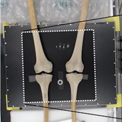
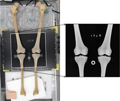
 An illustration of the x-ray procedure. The femora and tibiae of each skeleton were fixed on a board with a digital detector attached to it. Then, the tibiofemoral joint was reconstructed and placed in the center of the detector in a standard anatomical anteroposterior position. Sheets of styrofoam and bubble wrap were used to help adjust positioning. Finally, a 30-mm calibration disc and identification number of the specimen were placed onto the detector plate, and the radiograph was obtained. Radiographs were used instead of ordinary photographs as they were considered to capture both external and internal features of bone. Image courtesy of Legal Medicine through CC BY 4.0.
Preliminary work using new AI methods to estimate sex have used other skeletal sites, such as the cranium and pelvis, yet not many studies have explored AI-based sex estimation methods for the knee. To address the knowledge gap, the researchers established an AI deep-learning algorithm for estimating sex using digital knee x-rays from reconstructed knees of cadavers belonging to a Smithsonian skeletal research collection called the Terry Anatomical Collection.
The collection was created by Robert J. Terry (1871–1966) during his time as professor of anatomy at Washington University Medical School in St. Louis, MO, and includes 1,728 human skeletons. After Terry's death, the collection was donated to the museum for research.
The group in Finland obtained a total of 199 knee x-rays from 100 skeletons belonging to the collection. The sample included 46 male and 54 female cadavers, of whom 48 were classified as white and 52 as Black in associated documents. Mean age at death was 64.2 years with a range from 50 to 102 years.
The researchers uploaded the images and associated bone features in AIDeveloper, an open-source software with a variety of neural network architectures available for image classification. The images were split into three sets: One for training (70%), one for validation (15%), and another for testing (15%). The testing set was introduced in the final phase of the work.
Out of several algorithms generated by AIDeveloper, the deep-learning algorithm that performed the best achieved an overall testing accuracy of 90.3%. The model was able to classify all females and most males (78.6%) on the knee x-rays, according to the findings.
"The current findings suggest that deep learning algorithms based on knee radiography may have a future role in sex estimation, potentially alongside conventional methods," the researchers wrote.


